Tissue-Tek Genie® Advanced Staining Reagents
Quality is US – How we ensure optimal stain quality and scores for our antibodies
Laboratories are looking for staining slides with IHC assays that provide high sensitivity and high specificity to maximize diagnostic utility. Furthermore, they request consistent stain quality, slide after slide, especially important to always correctly detect low expressors. We devote our resources to produce reagents that always deliver optimal performance with a lot to lot consistency.
We have designed and implemented a straight forward process to develop robust assays with the following steps:
- Define the intended use and diagnostic utility of the assay
- Determine adequate control material
- Define acceptance criteria
- Identify best clone candidates
- Selection for each the best antigen retrieval
- Define for each the best dilution of the antibody and the incubation times for all the steps of the factory validated protocol
- Validate diagnostic performance internally and externally
Like NordiQC, a professional and scientific organization that promotes the quality of immunohistochemistry and expands its clinical use by arranging schemes for immunohistochemical proficiency testing, we publish the results from our validation studies on our own website called Advanced Staining Gallery to display assay performance scored optimal and enable laboratories to appreciate our daily work for you with full transparency.
We collaborate with a proficiency testing institute for external independent review and confirmation of successful fulfillment of the assessment and acceptance criteria, and for confirmation that the assay performance was scored optimal. As an example, please see the assessment / acceptance criteria for PMS2 applied and explained to the right.
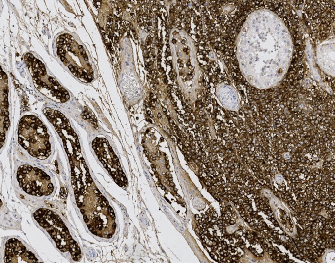
PMS2, like MSH6, MSH2 and MLH1, was scored optimal in the latest proficiency test runs of NordiQC (NordiCQ).
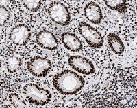
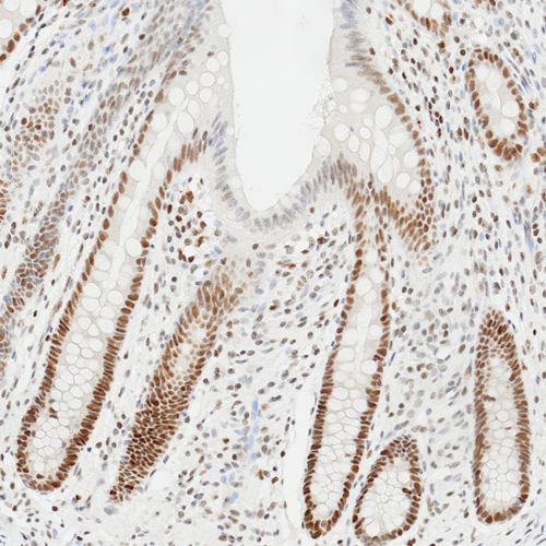
At least weak to moderte, distinct nuclear staining reaction of virtually all cells in the appendix.
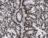
A moderate to strong, distinct nuclear staining raction of virtually all neoplastic cells in the colon adenocarcinoma.
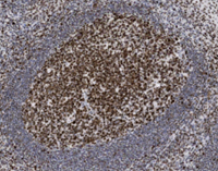
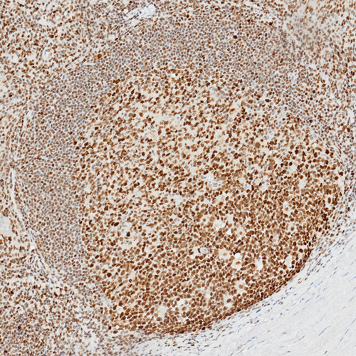
At least weak to moderate, distinct nuclear staining reaction of virtually all mantle zone B-cells and a moderate to strong, distinct nuclear staining reaction of the germinal center B-cells in the tonsil.
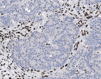
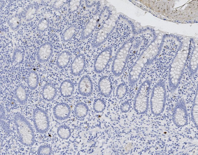
No nuclear staining reaction of the neoplastic cells in the colon adenocarcinomas, but a weak to moderate distinct nuclear staining reaction in the vast majority of other cells (stromal cells, lymphocytes etc.).
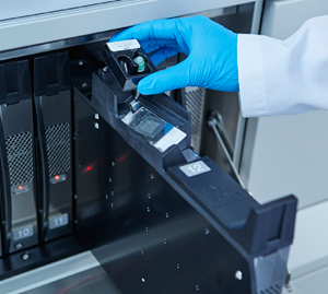
The innovative single-use Capsules make Genie the most convenient stainer on the market, eliminating batching and sorting of slides, and giving you the flexibility to load any slide, any stain, any time, any station on any Genie.
Benefit from using Capsules:
- Run cases or single slides without the need to batch slides by antibody
- Save time loading slides whenever a staining station is available
- Use for any slide volume of any biomarkers
The capsule is mounted to the Tissue-Tek Genie® Reagent Dispense Area (RDA) with its attached protocol RDA-Tag and both are loaded into an empty stain station.
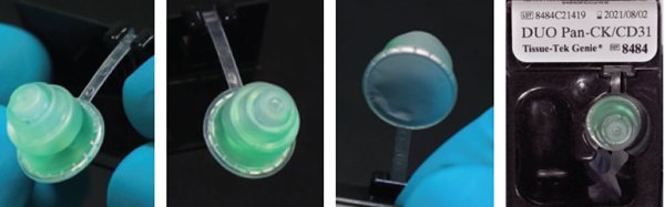
In summary, this innovative technology enables laboratories to quickly load slides and reagents for an IHC stain to gain continuous workflow with short and predictable TAT.
|
Product code |
Product name and quantity |
|---|---|
|
8637-K001 |
Tissue-Tek Genie® RDA-Tag Label Kit; 1 ribbon and 2 rolls of 2,400 RDA-Tag labels; 1 kit |
|
8616-G090 |
Tissue-Tek Genie® Reagent Dispense Area [RDA]; 90 RDAs; 1 box |
|
8618-G090 |
Tissue-Tek Genie® Reagent Dispense Area Tag [RDA-Tag]; 90 RDA-Tags; 1 box |
|
8636-K001 |
Tissue-Tek Genie® Slide Label Kit; 1 ribbon and 3 rolls of 3,000 slide labels; 1 kit |
|
8642-G001 |
Tissue-Tek Genie® Storage and Transport Tray, Blue; 1 unit |
|
8641-G001 |
Tissue-Tek Genie® Storage and Transport Tray, Red; 1 unit |
|
8217-G004 |
Tissue-Tek Genie® Waste Bottles |
|
8643-G001 |
Tissue-Tek Genie® Workflow Organizer; 1 unit |
|
9036 |
Tissue-Tek® SmartWrite® White Frosted Slides – Charged; 100/box; 10 boxes/case |
|
9046 |
Tissue-Tek® SmartWrite® Blue Frosted Slides – Charged, 100/box; 10 boxes/case |
|
9048 |
Tissue-Tek® SmartWrite® Green Frosted Slides – Charged, 100/box; 10 boxes/case |
|
9050 |
|
|
9052 |
Tissue-Tek® SmartWrite® Pink Frosted Slides – Charged, 100/box; 10 boxes/case |
|
9054 |
Tissue-Tek® SmartWrite® Yellow Frosted Slides – Charged, 100/box; 10 boxes/case |
|
9809-G006 |
Tissue-Tek Genie® CISH User-Fillable Cartridges for CISH Amplification Linker; 6 cartridges/box |
|
8610-K050 |
Genie® User-Fillable Capsule Sealing System; 1 kit |
|
8622-G006 |
Tissue-Tek Genie® User-Fillable Cartridge for Biomarker-Binding Reagent, 6 cartridges/box; 1 box |
|
8611-G002 |
Tissue-Tek Genie® User-Fillable Capsule Tray, 2/box; 1 box |
|
8615-G001 |
Tissue-Tek Genie® User-Fillable Capsule Sealer; 1 unit |
|
8619-K100 |
Tissue-Tek Genie® User-Fillable Capsules and Seals, 100 capsules, 130 seals; 1 kit |
Sakura Finetek offers 160 high quality primary antibodies and probes specifically developed to provide high sensitivity and specificity when used with the Tissue-Tek Genie Advanced Staining System. This ensures that users are provided with consistent, quality staining results and reproducibility that clinical laboratories have come to expect from Sakura Finetek IHC & ISH advanced staining products. These staining results were independently evaluated by a leading quality assurance organization to maximize diagnostic utility and provide optimal stain quality.
Discover our complete line of organ specific antibodies by clicking the buttons below.
Search Primary Antibodies
Tissue-Tek Genie® anti-Actin, Smooth Muscle
8292-M250: RTU, 250 tests, 1 cartridge; 1 unit
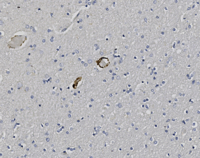
Tissue-Tek Genie® anti-ALK (Heme)
8350-M250: RTU, 250 tests, 1 cartridge; 1 unit
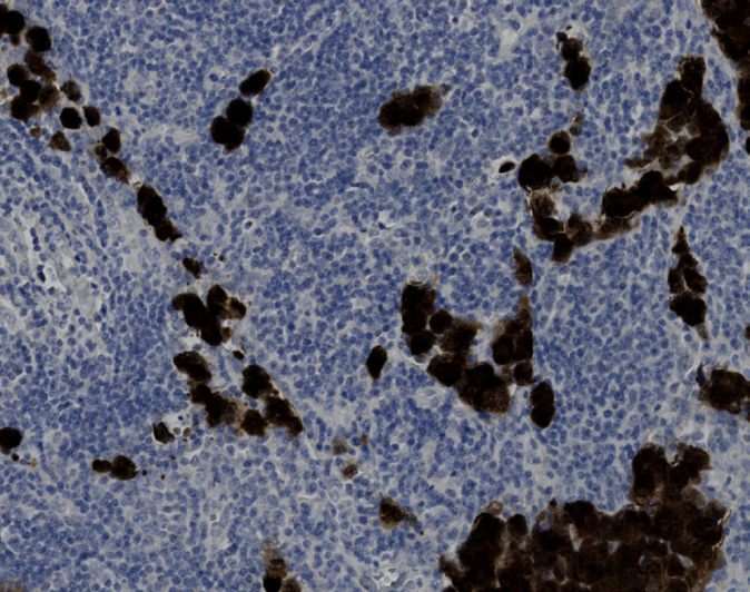
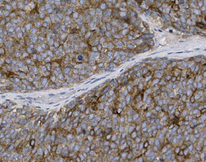
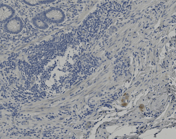
Tissue-Tek Genie® anti-AMACR
8291-M250: RTU, 1 cartridge, 250 tests; 1 unit
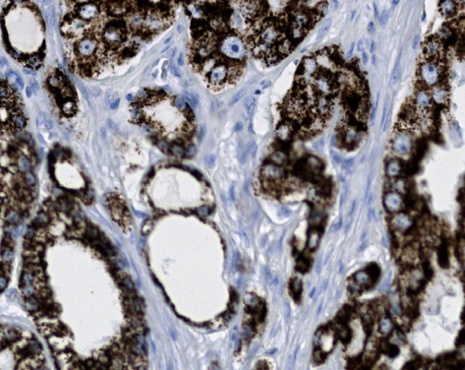
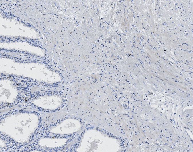
Tissue-Tek Genie® anti-BCL2
8459-M250: RTU, 1 cartridge, 250 tests; 1 unit
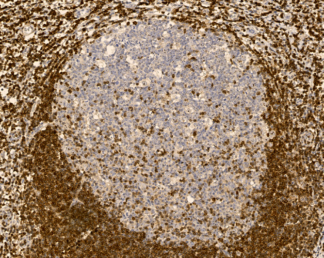
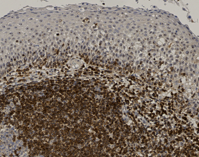
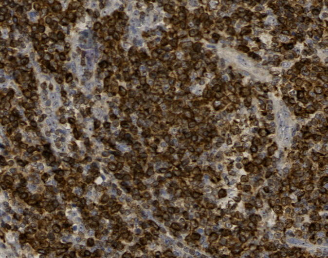
Tissue-Tek Genie® anti-BCL6
8461-M250: RTU, 1 cartridge, 250 tests; 1 unit
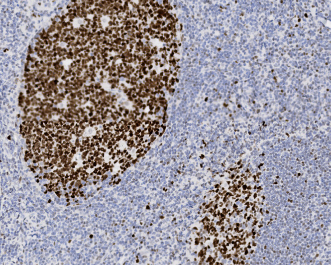
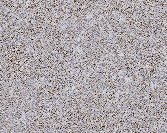
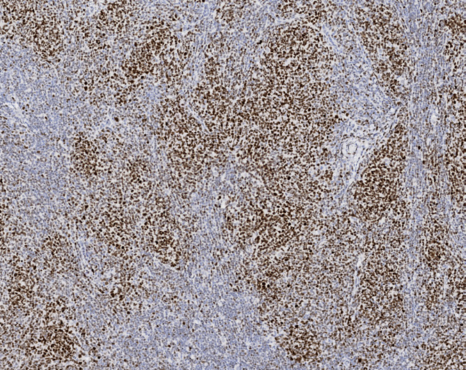
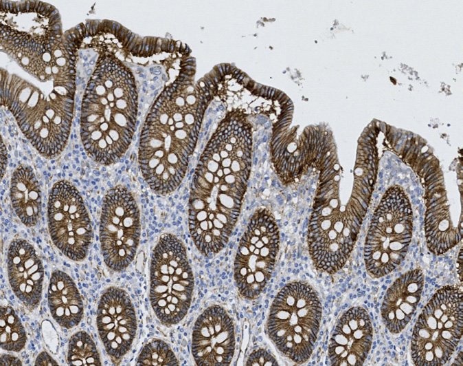
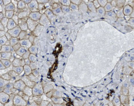
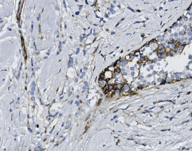
Tissue-Tek Genie® anti-beta-Catenin
8261-M250: RTU, 250 tests, 1 cartridge; 1 unit
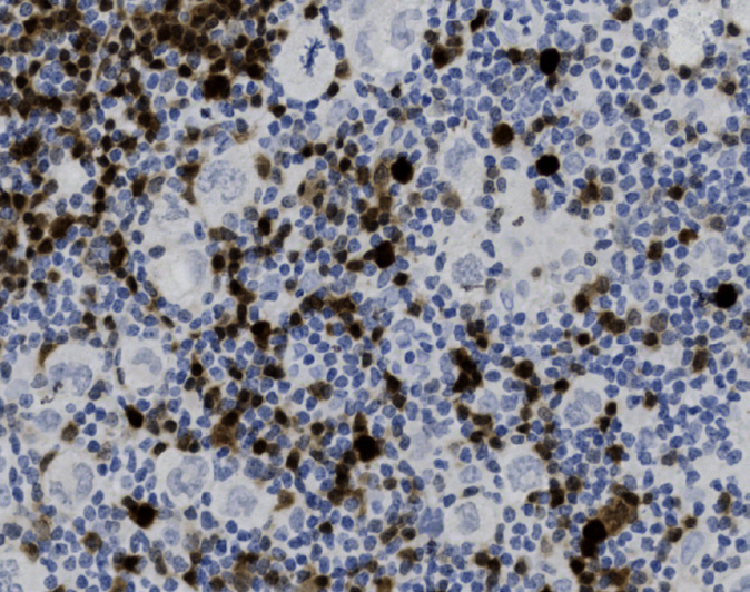
Tissue-Tek Genie® anti-BOB.1
8462-M250: RTU, 250 tests, 1 cartridge; 1 unit
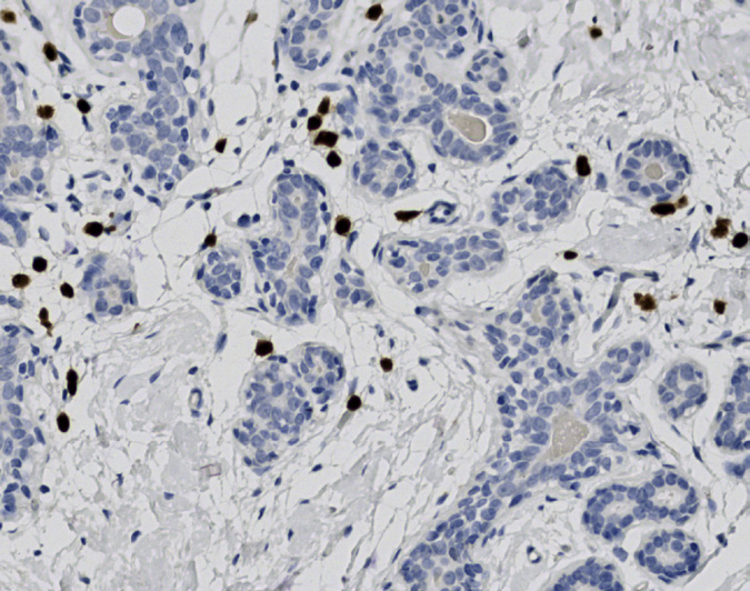
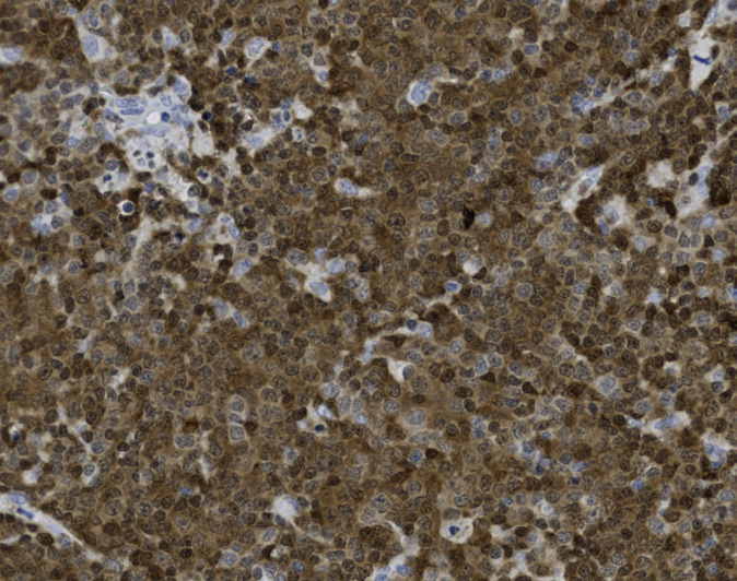
Tissue-Tek Genie® anti-c-Myc
8332-M250: RTU, 250 tests, 1 cartridge; 1 unit
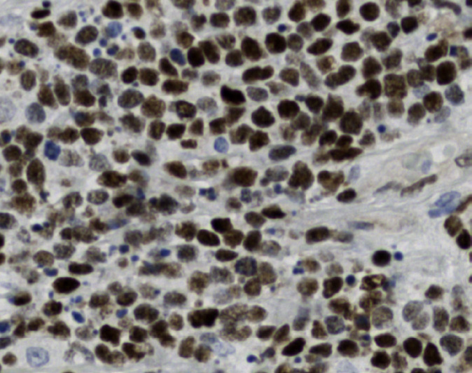
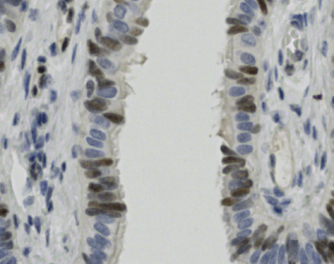
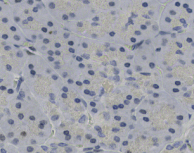
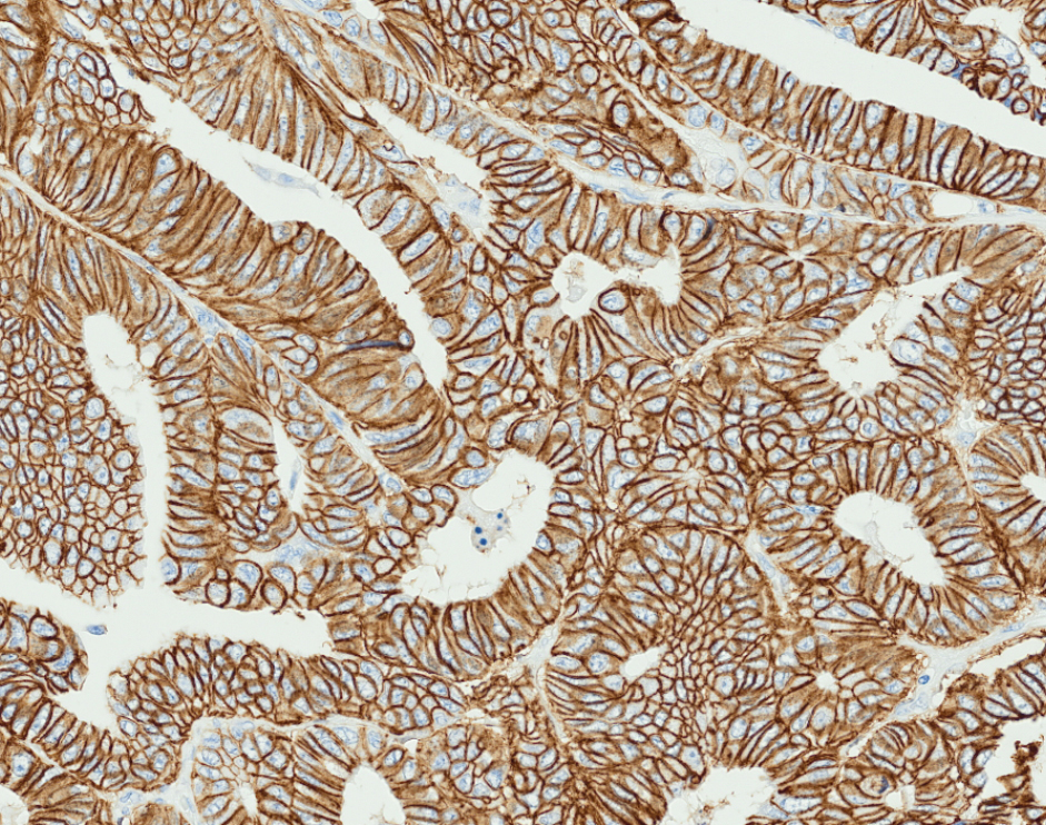
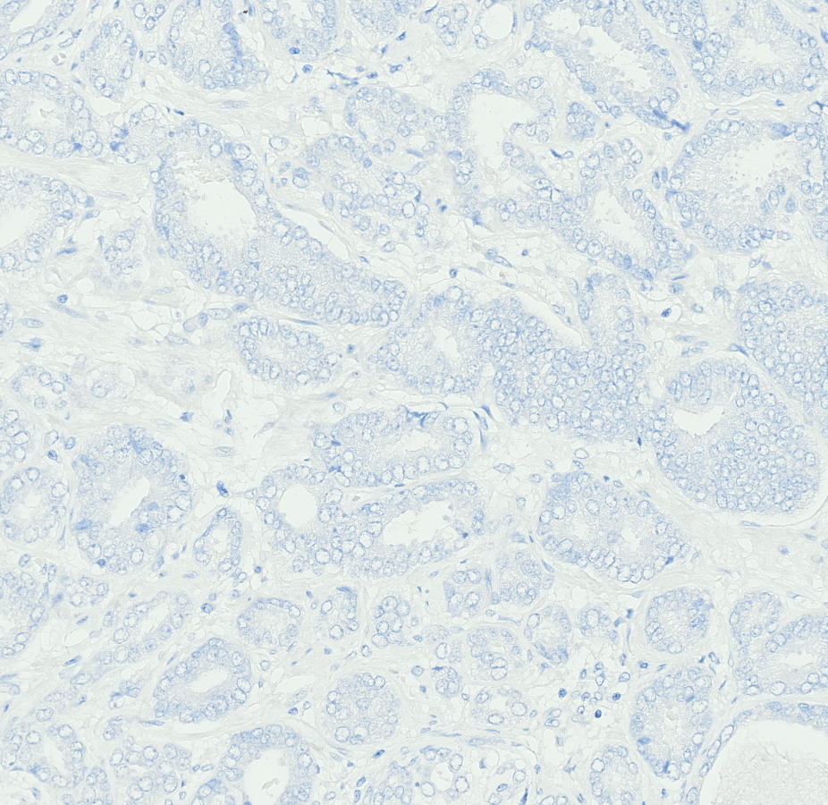
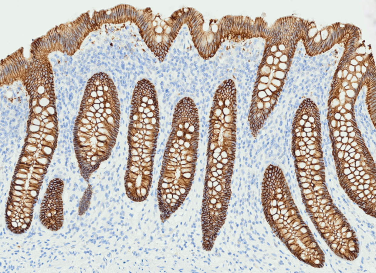
Tissue-Tek Genie® anti-Cadherin 17
8364-M250: RTU, 250 tests, 1 cartridge; 1 unit
Cadherin 17 (CDH17), also known as liver-intestinal cadherin (LIcadherin), is a transmembrane glycoprotein involved in cell adhesion.
Tissue-Tek Genie® anti-Caldesmon
8345-M250: RTU, 250 tests, 1 cartridge; 1 unit
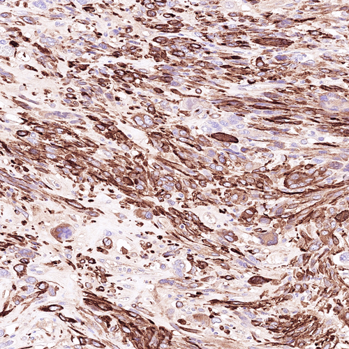
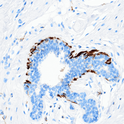
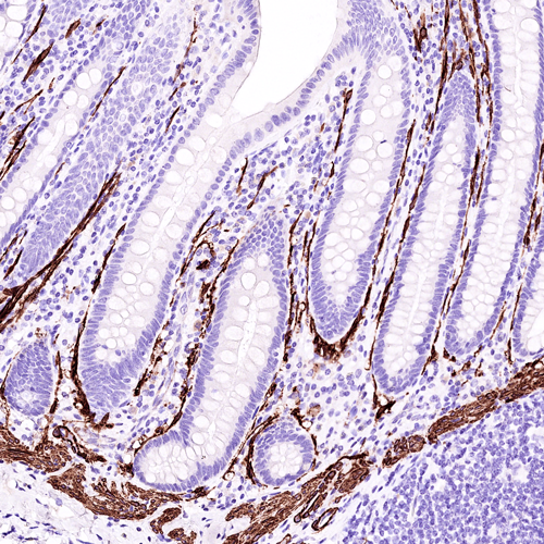
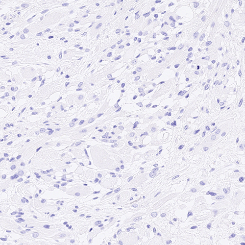
The Tissue-Tek Genie® anti-Caldesmon Mouse Monoclonal Antibody [ZM79] is designed to qualitatively detect Caldesmon protein in formalin-fixed, paraffin-embedded (FFPE) human tissue specimen sections by immunohistochemistry (IHC) staining on the automated Tissue-Tek Genie® Advanced Staining System.
This device functions as an aid for diagnosis and shall be used by a qualified pathologist with a panel of other antibodies to identify tumors with smooth muscle cell differentiation.
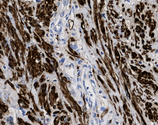
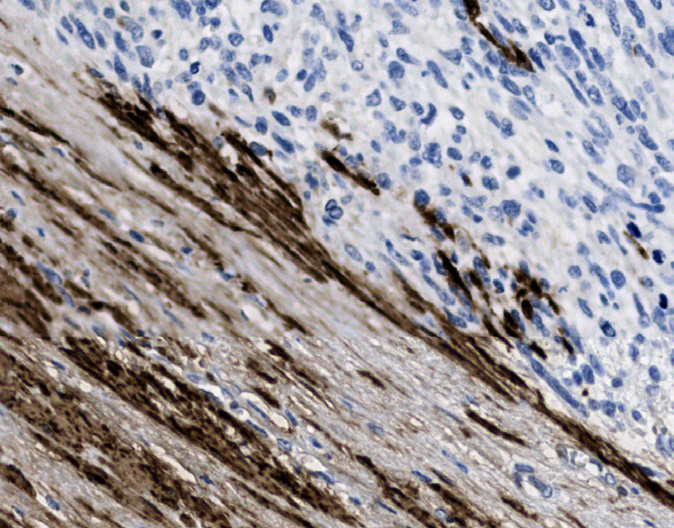
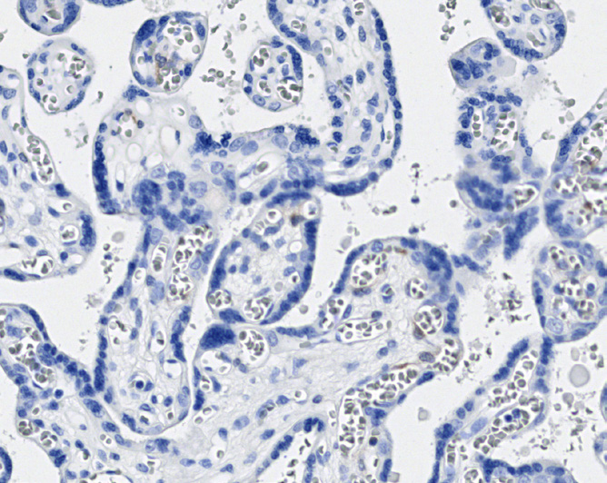
Tissue-Tek Genie® anti-Calponin
8310-M250: RTU, 1 cartridge, 250 tests; 1 unit
Tissue-Tek Genie® anti-Calretinin
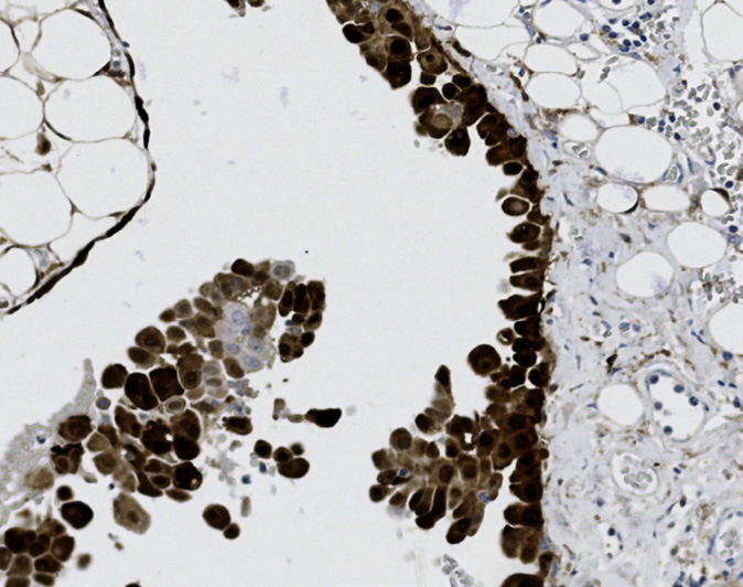
8522-M250: RTU, 250 tests, 1 cartridge; 1 unit
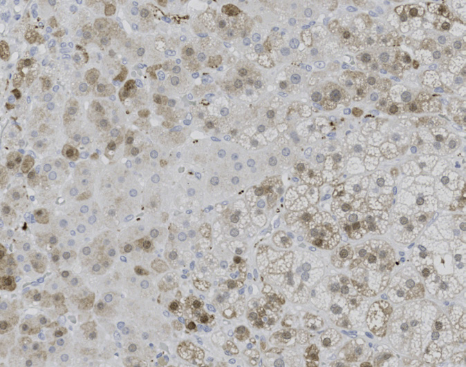
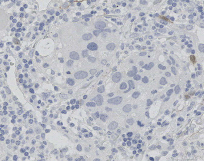
Tissue-Tek Genie® anti-CD1a
8246-M250: RTU, 1 cartridge, 250 tests; 1 unit
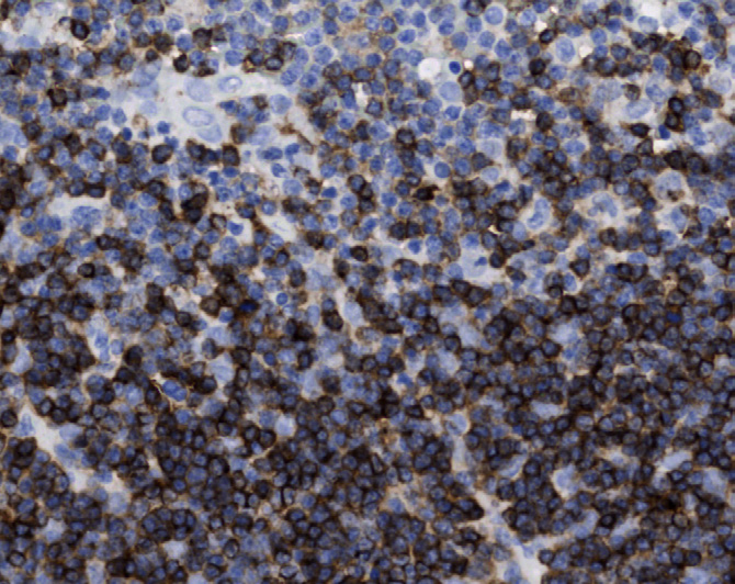
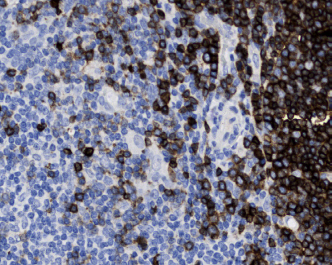
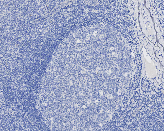
Tissue-Tek Genie® anti-CD2
8255-M250: RTU, 1 cartridge, 250 tests; 1 unit
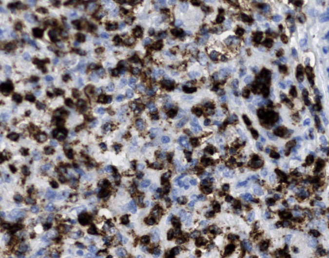
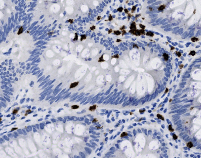
Tissue-Tek Genie® anti-CD3 [EP177]
8280-M250: RTU, 250 tests, 1 cartridge; 1 unit
The Tissue-Tek Genie® anti-CD3 Rabbit Monoclonal Antibody [EP177] is designed to qualitatively detect CD3 protein in formalin-fixed, paraffin-embedded (FFPE) human tissue specimen sections by immunohistochemistry (IHC) staining on the automated Tissue-Tek Genie® Advanced Staining System.
This device functions as an aid for diagnosis and shall be used by a qualified pathologist with a panel of other antibodies to identify T-cell neoplasms and highlight T-cell populations within a variety of tissues.
![Tissue-Tek Genie® anti-CD3 [EP177]](https://www.sakuraus.com/wp-content/uploads/2026/10/cd3-ep177-1.png)
![Tissue-Tek Genie® anti-CD3 [EP177] - Additional Image 1](https://www.sakuraus.com/wp-content/uploads/2026/10/cd3-ep177-2.png)
![Tissue-Tek Genie® anti-CD3 [EP177] - Additional Image 2](https://www.sakuraus.com/wp-content/uploads/2026/10/cd3-ep177-3.png)
![Tissue-Tek Genie® anti-CD3 [EP177] - Additional Image 3](https://www.sakuraus.com/wp-content/uploads/2026/10/cd3-ep177-4.png)
Tissue-Tek Genie® anti-CD3 [ZM45]
8344-M250: RTU, 250 tests, 1 cartridge; 1 unit
The Tissue-Tek Genie® anti-CD3 Mouse Monoclonal Antibody [ZM45] is designed to qualitatively detect CD3 protein in formalin-fixed, paraffin-embedded (FFPE) human tissue specimen sections by immunohistochemistry (IHC) staining on the automated Tissue-Tek Genie® Advanced Staining System.
This device functions as an aid for diagnosis and shall be used by a qualified pathologist with a panel of other antibodies to identify T-cell neoplasms and highlight T-cell populations within a variety of tissues.
![Tissue-Tek Genie® anti-CD3 [ZM45]](https://www.sakuraus.com/wp-content/uploads/2026/10/cd3-zm45-1.png)
![Tissue-Tek Genie® anti-CD3 [ZM45] - Additional Image 1](https://www.sakuraus.com/wp-content/uploads/2026/10/cd3-zm45-2.png)
![Tissue-Tek Genie® anti-CD3 [ZM45] - Additional Image 2](https://www.sakuraus.com/wp-content/uploads/2026/10/cd3-zm45-3.png)
![Tissue-Tek Genie® anti-CD3 [ZM45] - Additional Image 3](https://www.sakuraus.com/wp-content/uploads/2026/10/cd3-zm45-4.png)
Tissue-Tek Genie® anti-CD4
8226-M250: RTU, 1 cartridge, 250 tests; 1 unit
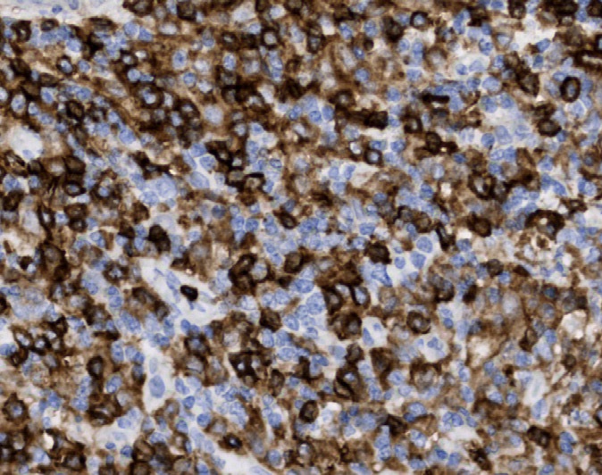
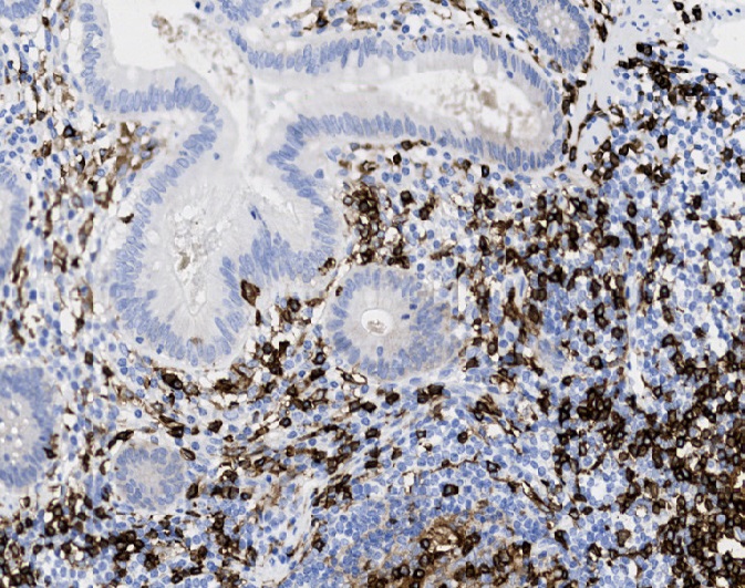
Tissue-Tek Genie® anti-CD5
8249-M250: RTU, 250 tests, 1 cartridge; 1 unit
CD5 is a transmembrane protein expressed on all mature T-cells and subsets of B-cells found in the follicular mantle zones, bone marrow, and peripheral blood.
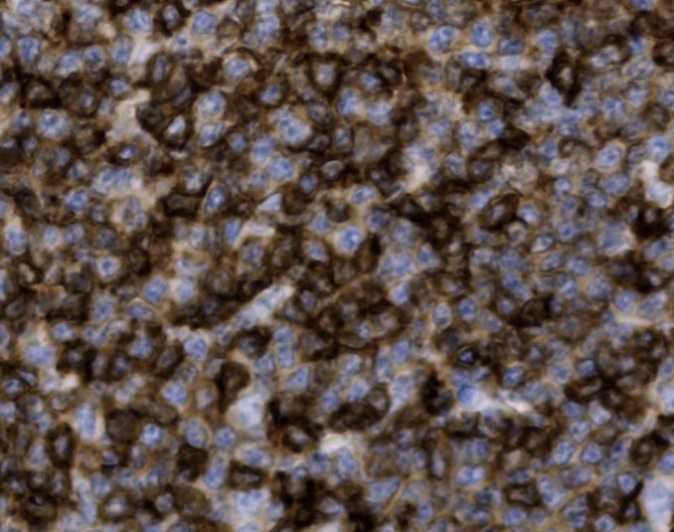
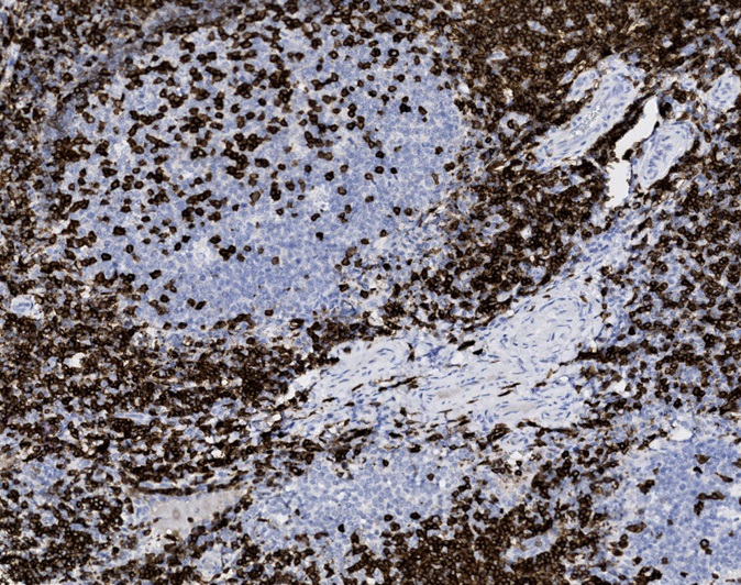
Tissue-Tek Genie® anti-CD7
8251-M250: RTU, 1 cartridge, 250 tests; 1 unit
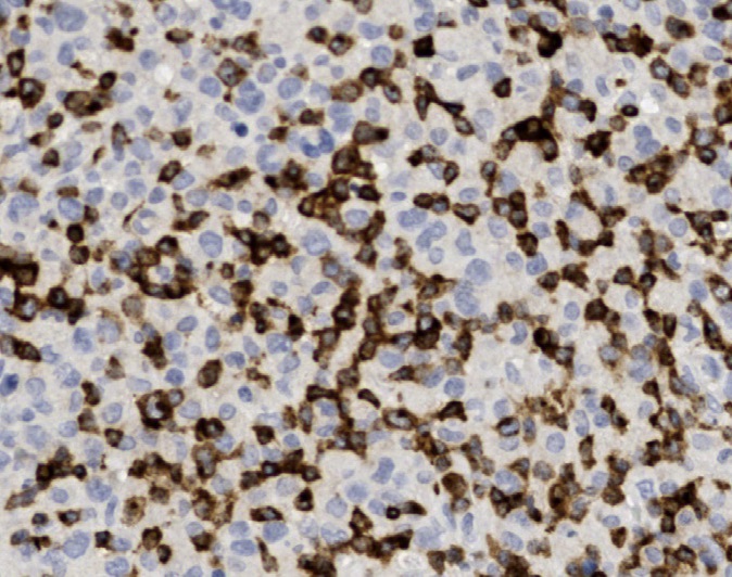
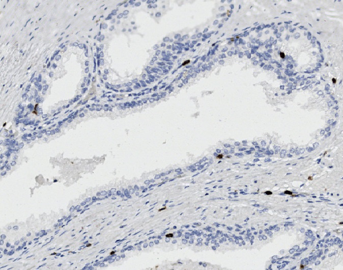
Tissue-Tek Genie® anti-CD8
8252-M250: RTU, 1 cartridge, 250 tests; 1 unit
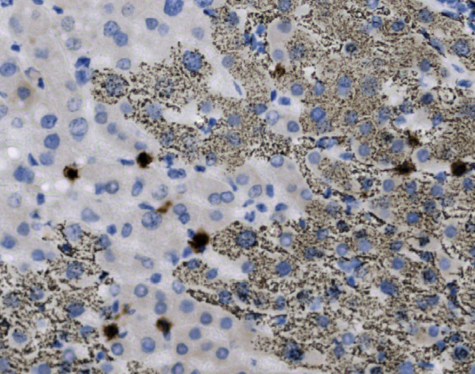
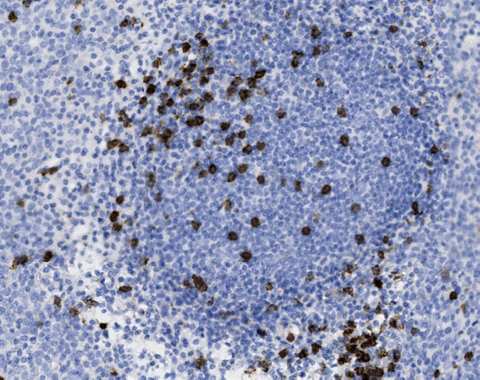
Tissue-Tek Genie® anti-CD10 [QR021]
8386-M250: RTU, 250 tests, 1 cartridge; 1 unit
The Tissue-Tek Genie® anti-CD10 Rabbit Monoclonal Antibody [QR021] is designed to qualitatively detect CD10 protein in formalin-fixed, paraffin-embedded (FFPE) human tissue specimen sections by immunohistochemistry (IHC) staining on the automated Tissue-Tek Genie® Advanced Staining System.
This device functions as an aid for diagnosis and shall be used by a qualified pathologist with a panel of other antibodies to classify a subset of B-cell lymphomas such as diffuse large B-cell lymphoma (DLBCL), follicular lymphoma and Burkitt lymphoma, and renal cell carcinoma.
![Tissue-Tek Genie® anti-CD10 [QR021]](https://www.sakuraus.com/wp-content/uploads/2026/10/cd10-3.png)
![Tissue-Tek Genie® anti-CD10 [QR021] - Additional Image 1](https://www.sakuraus.com/wp-content/uploads/2026/10/cd10-1.png)
![Tissue-Tek Genie® anti-CD10 [QR021] - Additional Image 2](https://www.sakuraus.com/wp-content/uploads/2026/10/cd10-2.png)
![Tissue-Tek Genie® anti-CD10 [QR021] - Additional Image 3](https://www.sakuraus.com/wp-content/uploads/2026/10/cd10-4.png)
Tissue-Tek Genie® anti-CD13
8503-M250: RTU, 250 tests, 1 cartridge; 1 unit
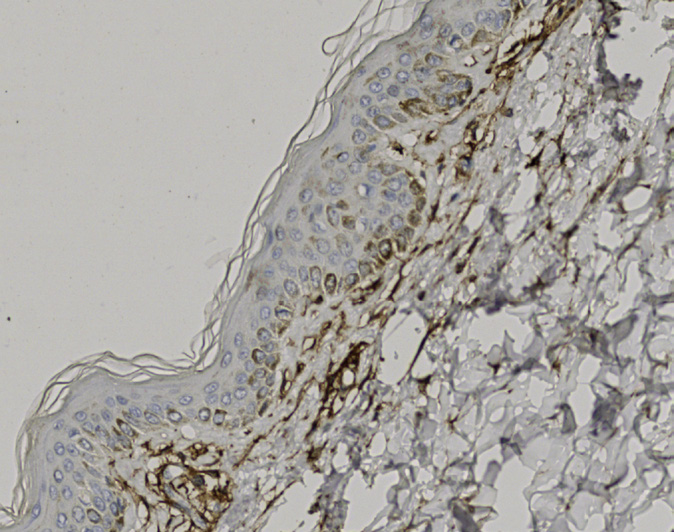
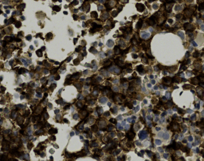
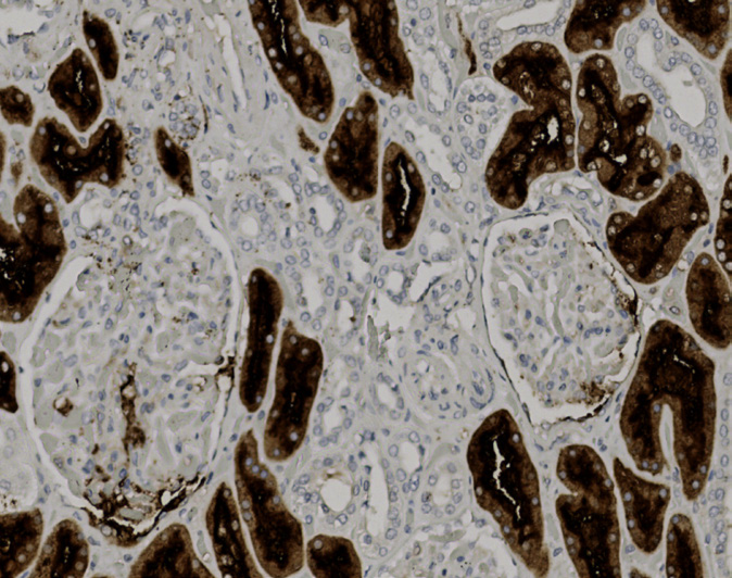
Tissue-Tek Genie® anti-CD14
8321-M250: RTU, 250 tests, 1 cartridge; 1 unit
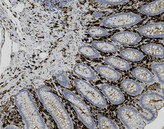
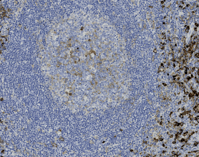
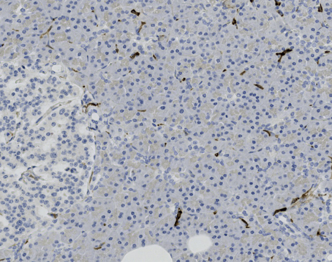
Tissue-Tek Genie® anti-CD15
8256-M250: RTU, 250 tests, 1 cartridge; 1 unit
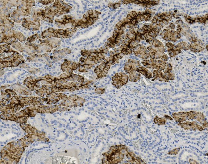
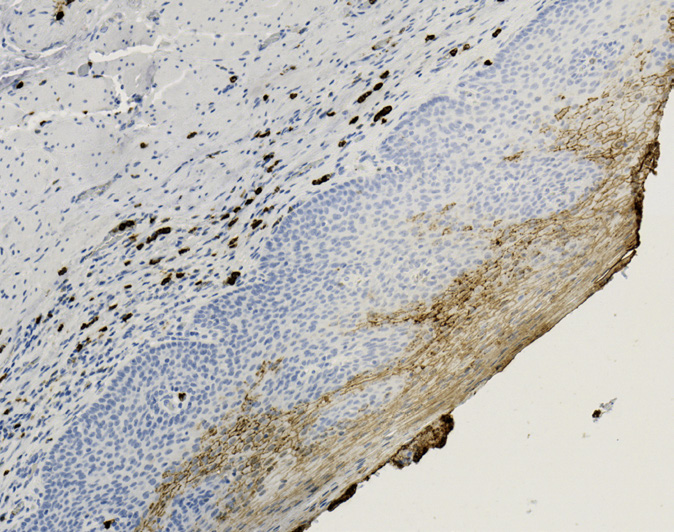
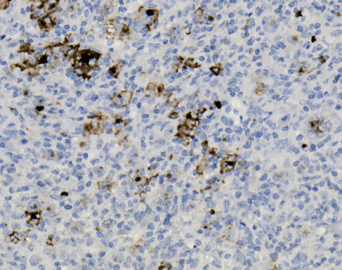
Tissue-Tek Genie® anti-CD19
8516-M250: RTU, 250 tests, 1 cartridge; 1 unit
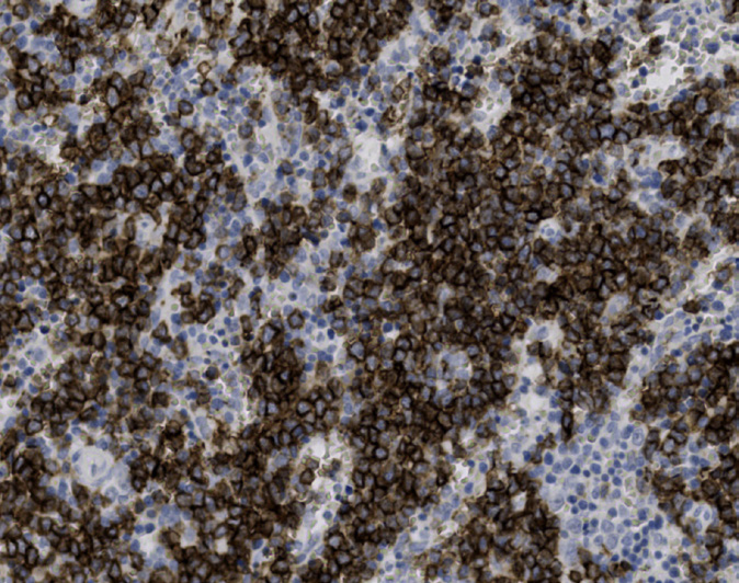
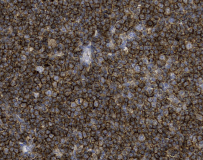
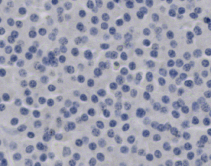
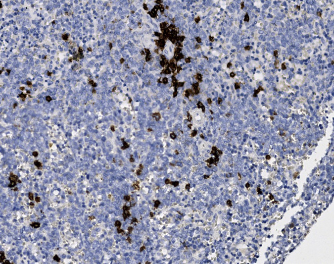
Tissue-Tek Genie® anti-CD20
8259-M250: RTU, 1 cartridge, 250 tests; 1 unit
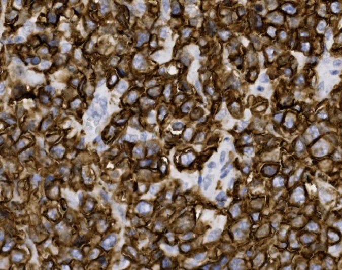
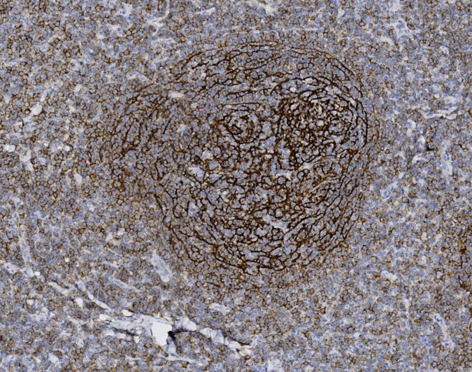
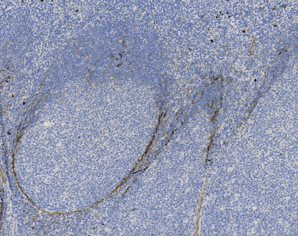
Tissue-Tek Genie® anti-CD21
8260-M250: RTU, 250 tests, 1 cartridge; 1 unit
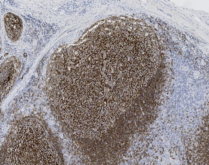
Tissue-Tek Genie® anti-CD23
8262-M250: RTU, 1 cartridge, 250 tests; 1 unit
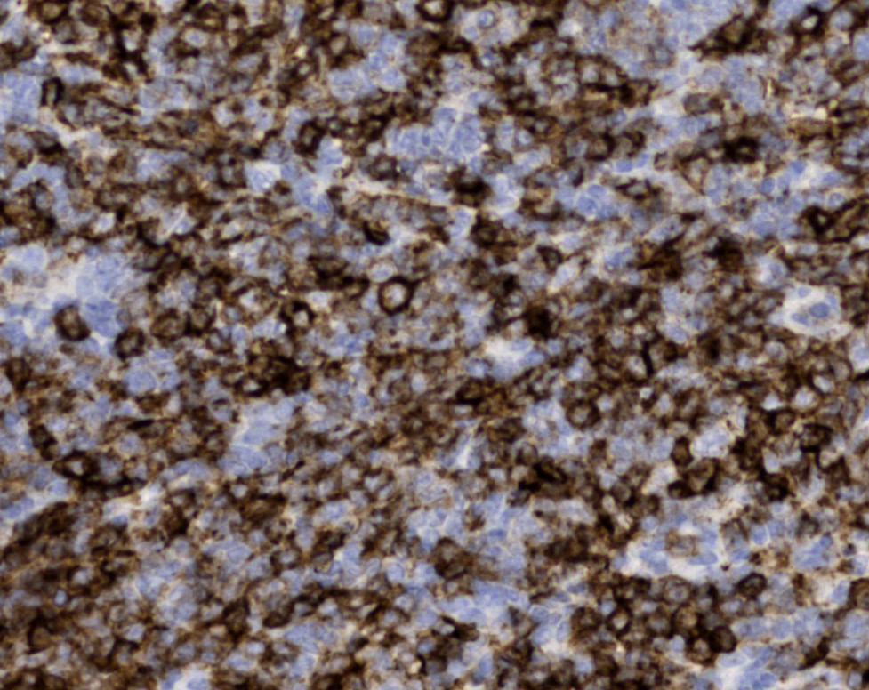
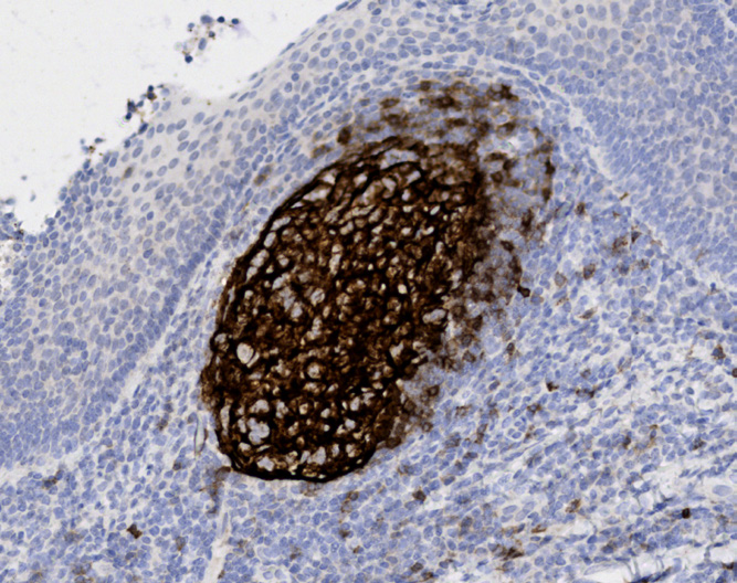
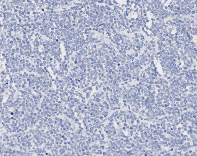
Tissue-Tek Genie® anti-CD30
8265-M250: RTU, 1 cartridge, 250 tests; 1 unit
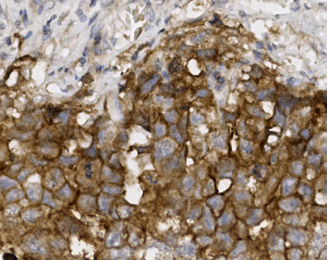
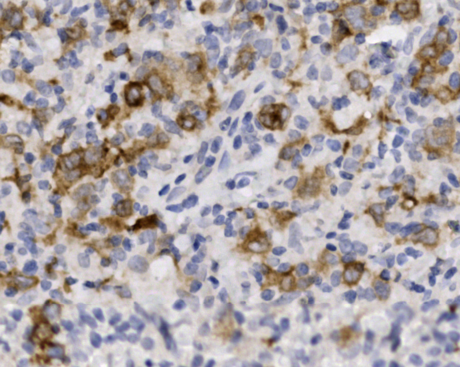
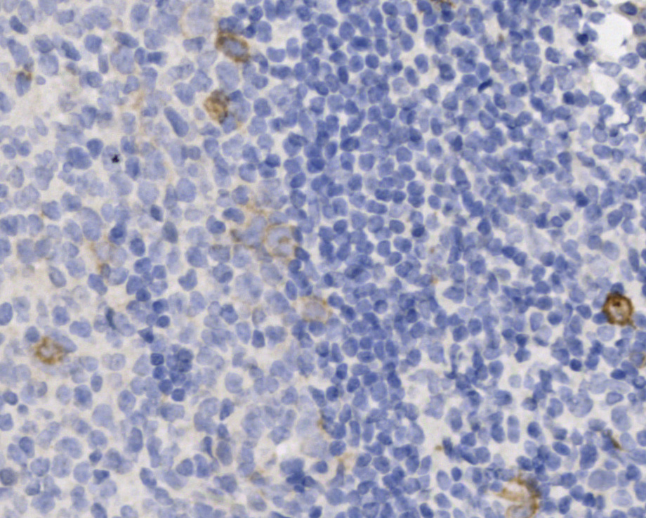
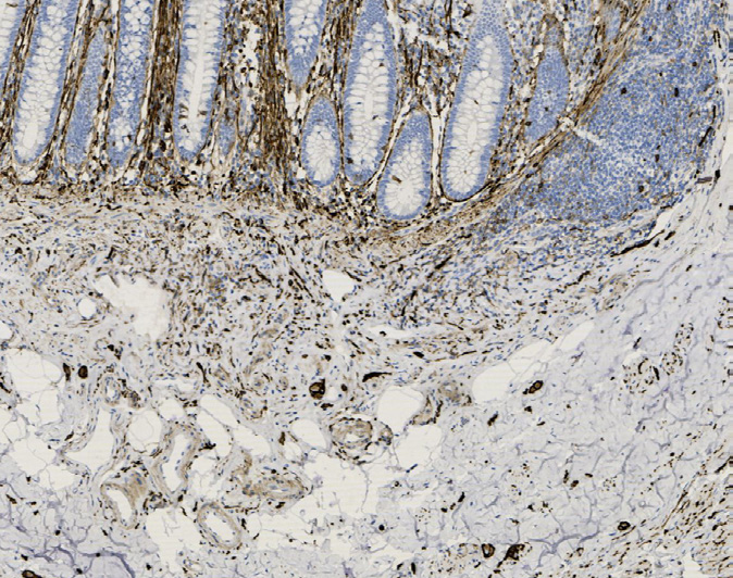
Tissue-Tek Genie® anti-CD31
8282-M250: RTU, 1 cartridge, 250 tests; 1 unit
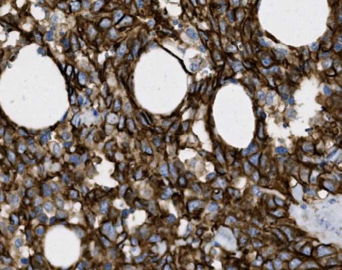
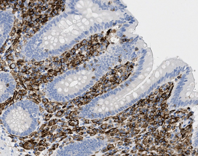
Tissue-Tek Genie® anti-CD34
8268-M250: RTU, 1 cartridge, 250 tests; 1 unit
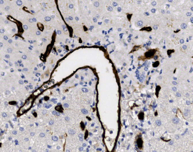
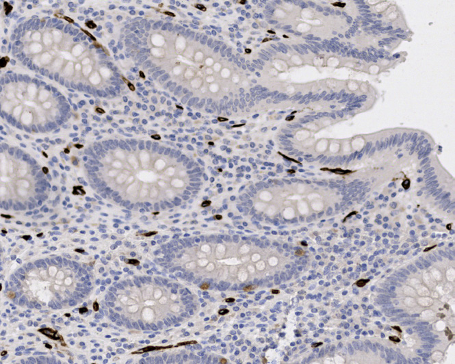
Positive tissue
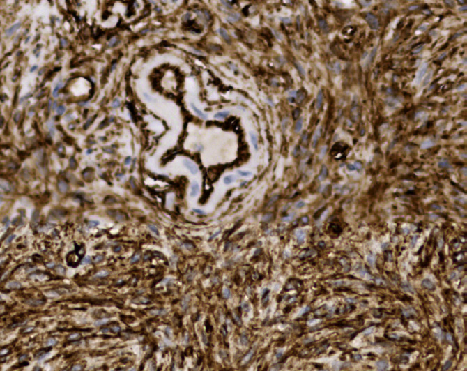
Tissue-Tek Genie® anti-CD38
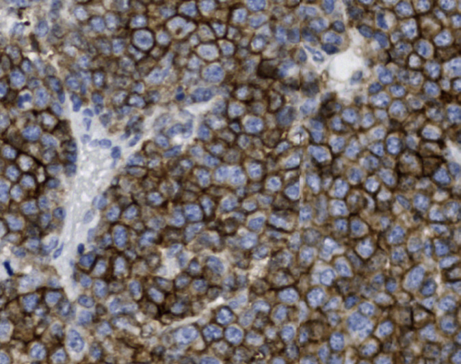
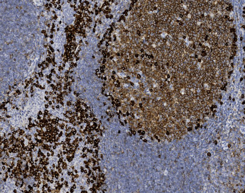
8277-M250: RTU, 250 tests, 1 cartridge; 1 unit
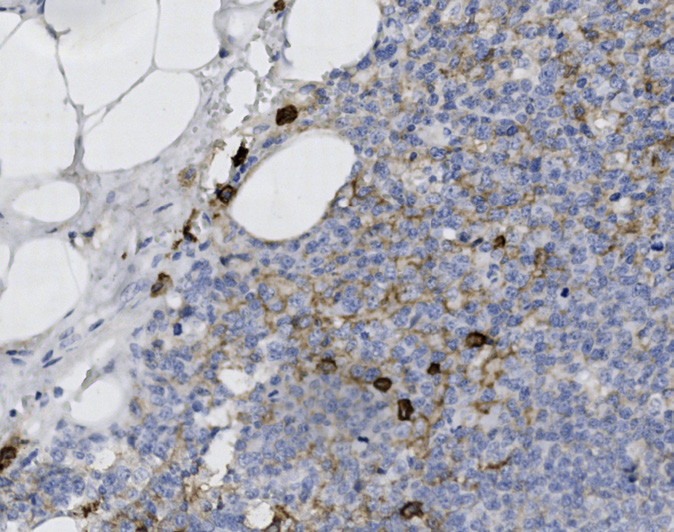
Tissue-Tek Genie® anti-CD43
8279-M250: RTU, 250 tests, 1 cartridge; 1 unit
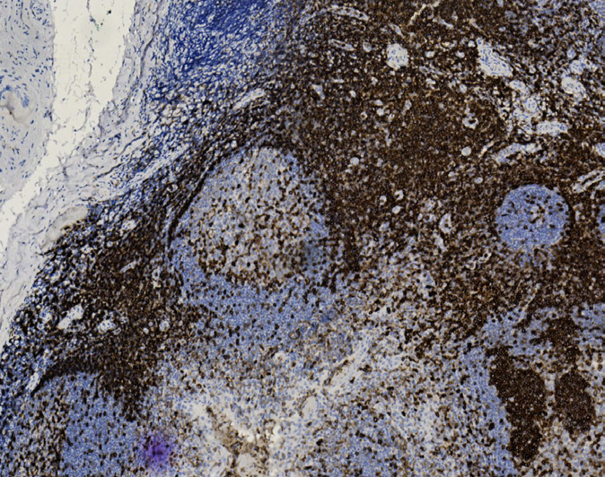
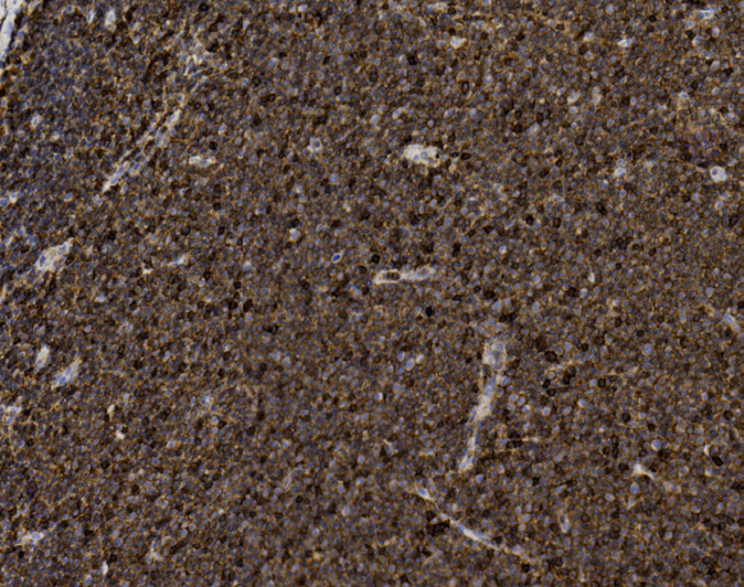
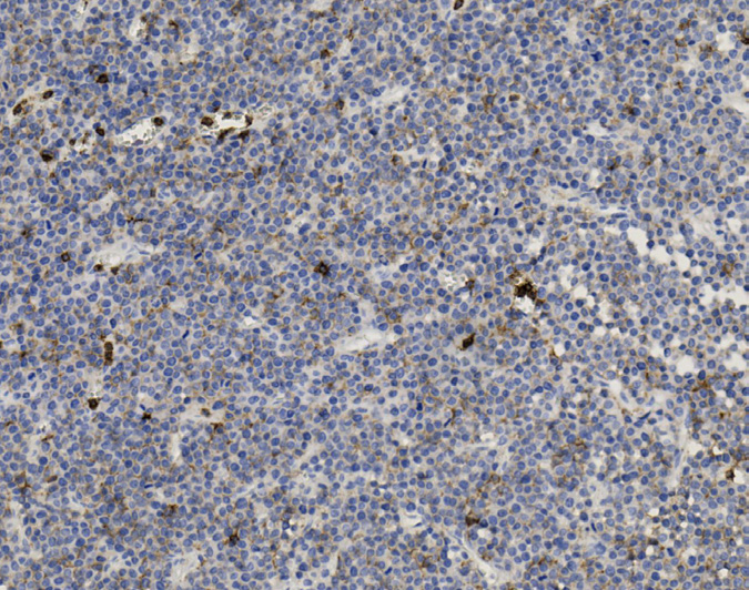
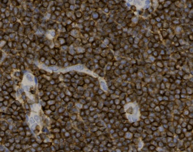
Tissue-Tek Genie® anti-CD45
8271-M250: RTU, 1 cartridge, 250 tests; 1 unit
Tissue-Tek Genie® anti-CD56
8274-M250: RTU, 250 tests, 1 cartridge; 1 unit
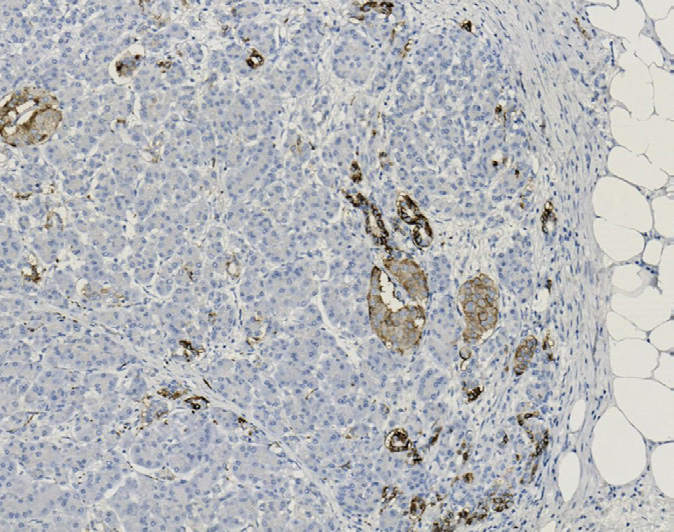
Tissue-Tek Genie® anti-CD57
8338-M250: RTU, 250 tests, 1 cartridge; 1 unit
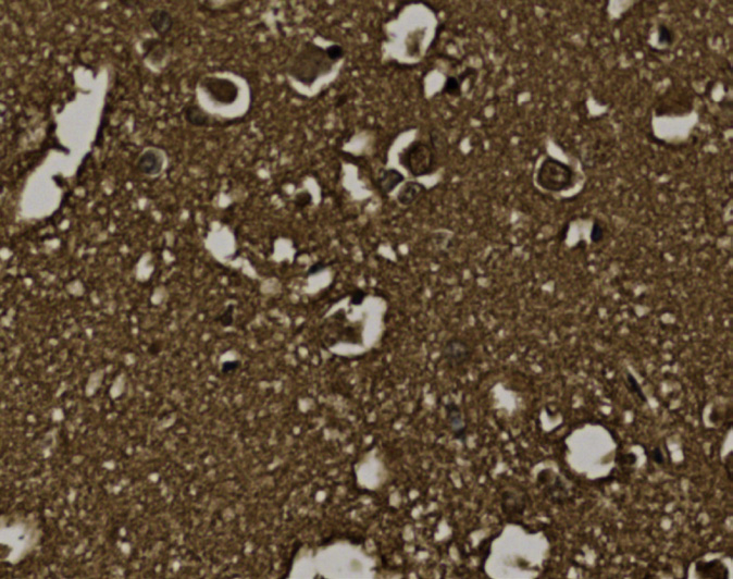
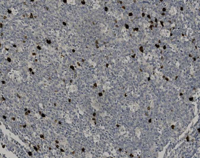
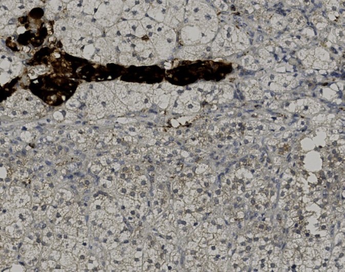
Tissue-Tek Genie® anti-CD61
8518-M250: RTU, 250 tests, 1 cartridge; 1 unit
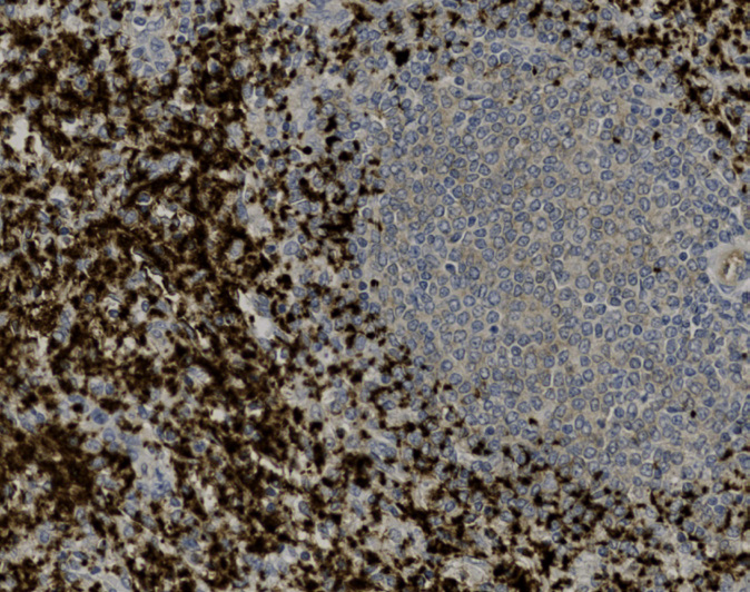
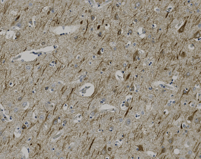
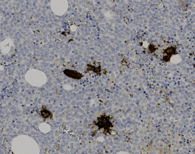
Tissue-Tek Genie® anti-CD63
8283-M250: RTU, 250 tests, 1 cartridge; 1 unit
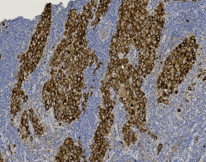
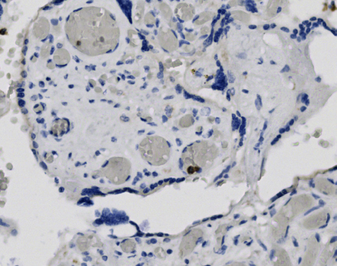
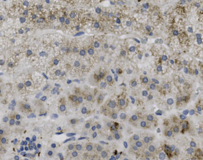
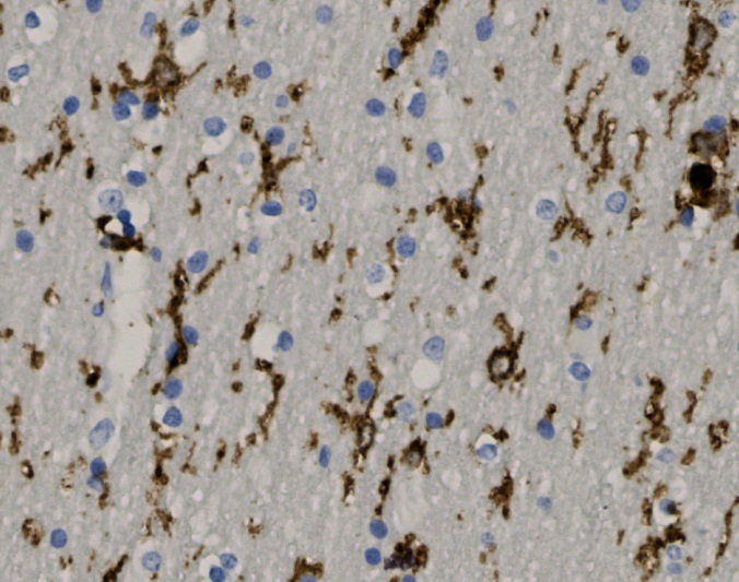
Tissue-Tek Genie® anti-CD68
8275-M250: RTU, 1 cartridge, 250 tests; 1 unit
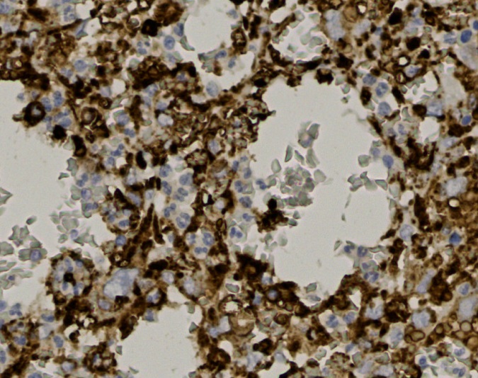
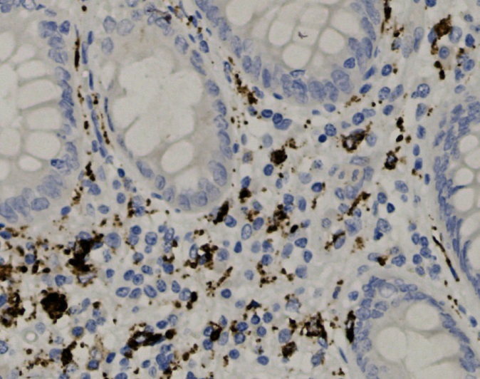
Tissue-Tek Genie® anti-CD71
8322-M250: RTU, 250 tests, 1 cartridge; 1 unit
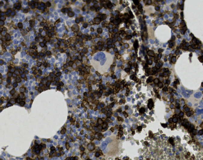
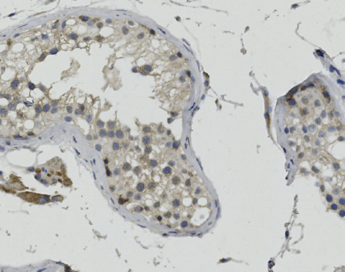
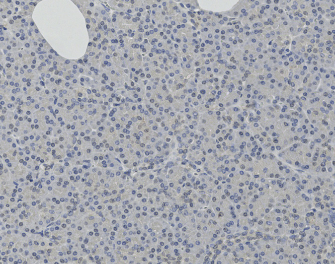
Tissue-Tek Genie® anti-CD79a
8278-M250: RTU, 1 cartridge, 250 tests; 1 unit
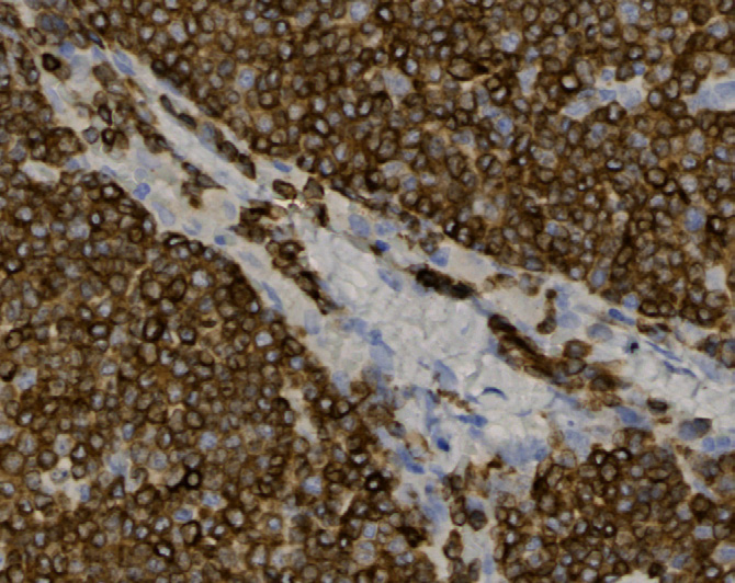
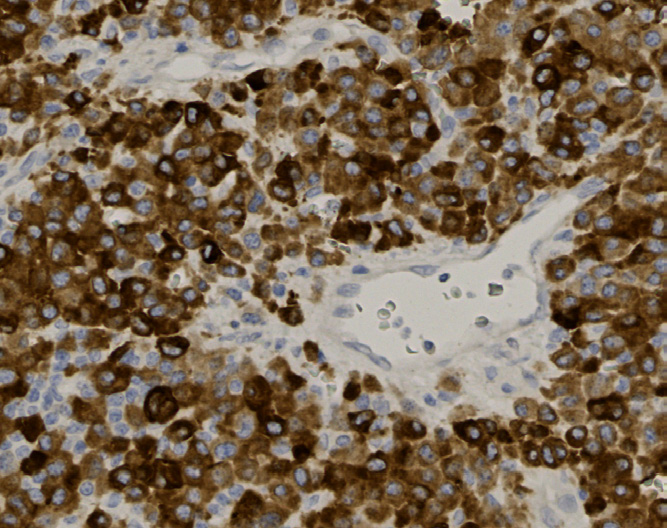
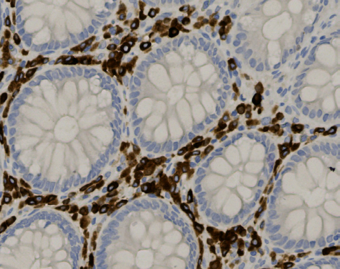
Tissue-Tek Genie® anti-CD117 (c-kit)
8267-M250: RTU, 250 tests, 1 cartridge; 1 unit
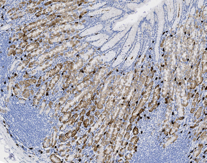
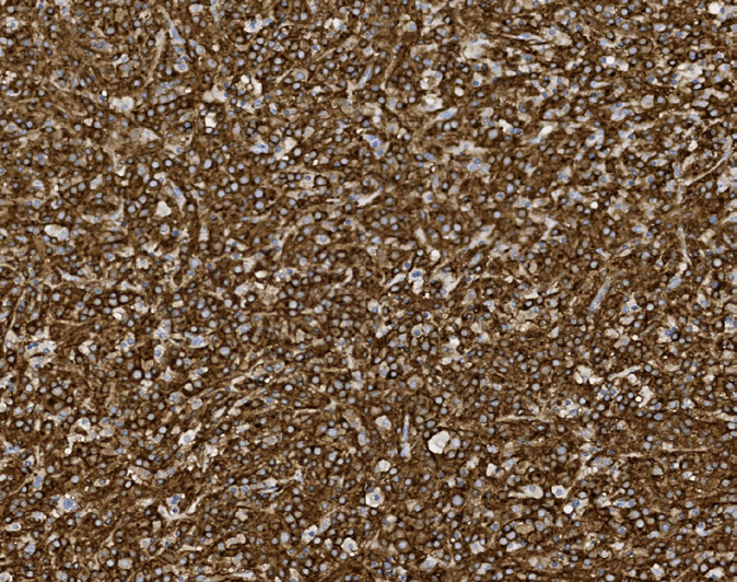
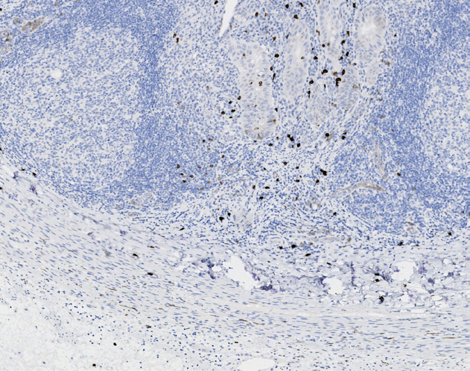
Tissue-Tek Genie® anti-CD138
8241-M250: RTU, 250 tests, 1 cartridge; 1 unit
of epithelial cells are CD138 positive.
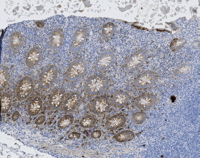
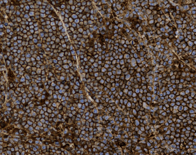
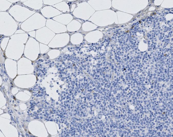
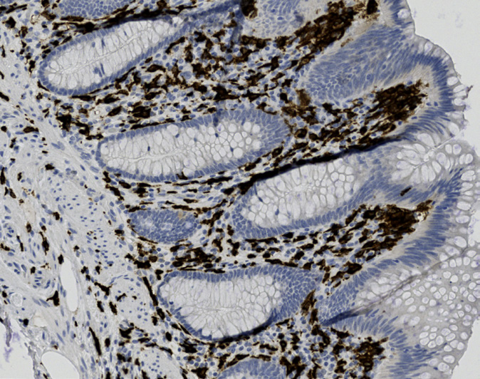
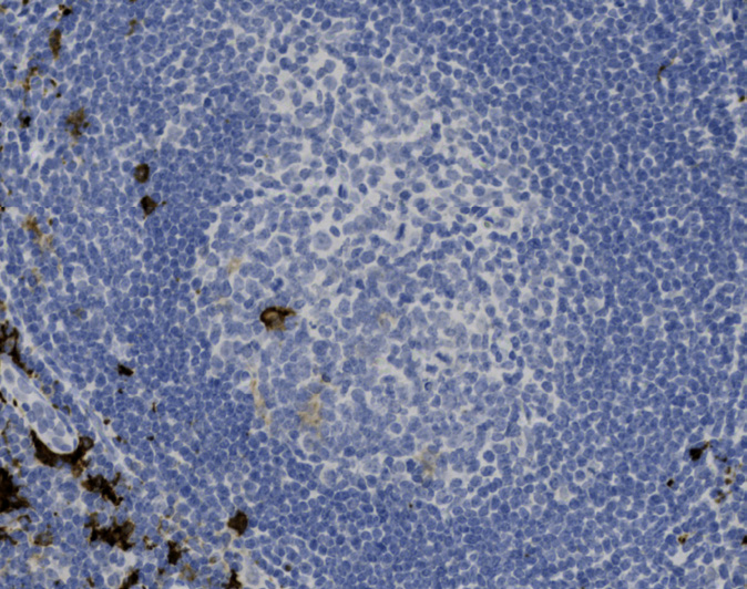
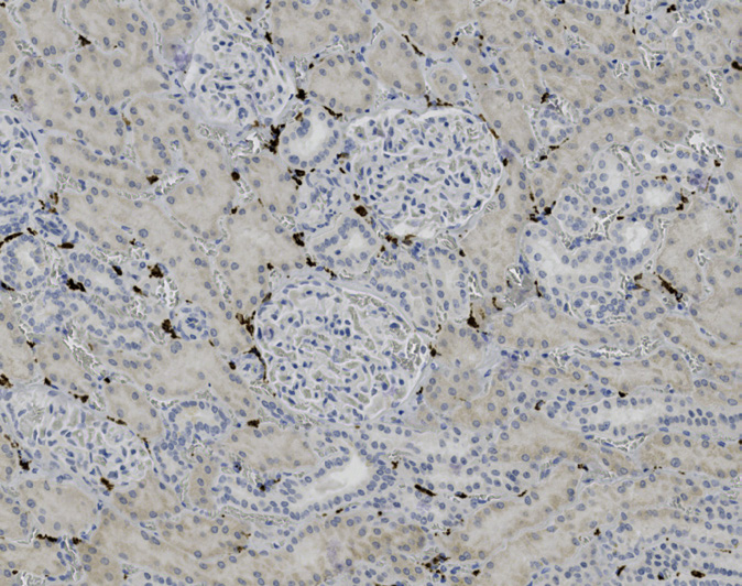
Tissue-Tek Genie® anti-CD163
8245-M250: RTU, 250 tests, 1 cartridge; 1 unit
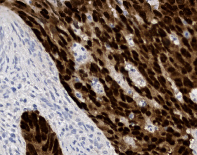
Tissue-Tek Genie® anti-CDX2
8285-M250: RTU, 1 cartridge, 250 tests; 1 unit
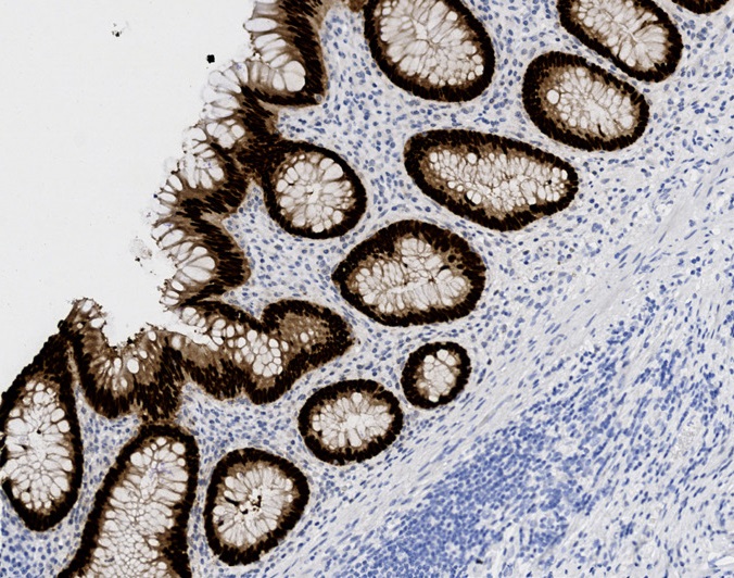
Tissue-Tek Genie® anti-CEA
8225-M250: RTU, 1 cartridge, 250 tests; 1 unit
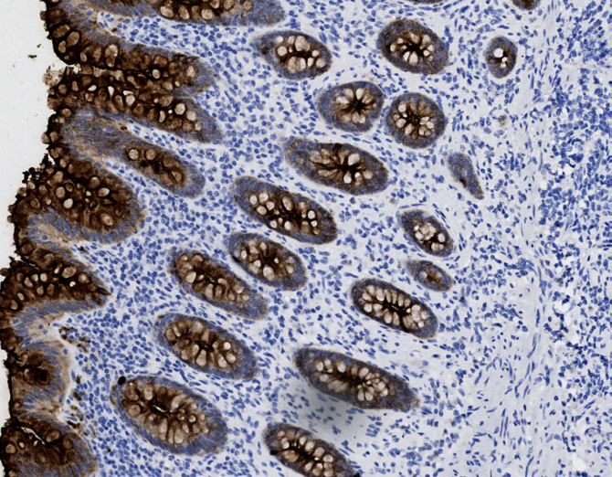
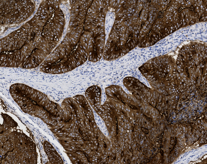
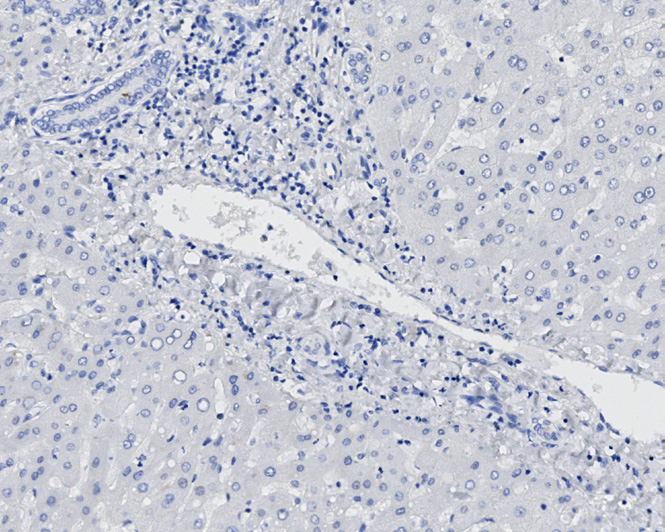
Tissue-Tek Genie® anti-Chromogranin A
8286-M250: RTU, 1 cartridge, 250 tests; 1 unit
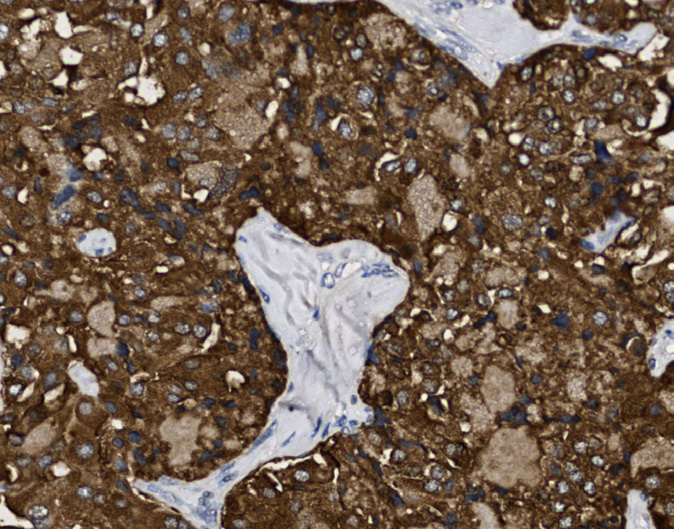
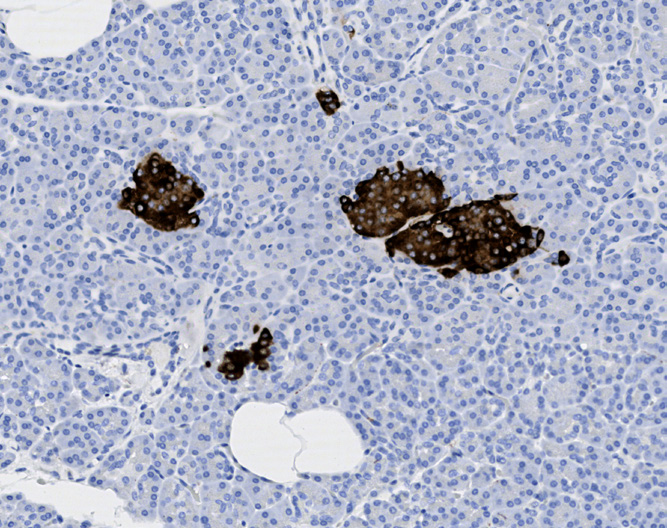
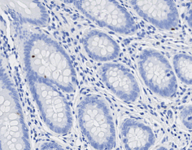
Tissue-Tek Genie® anti-Cyclin D1
8469-M250: RTU, 1 cartridge, 250 tests; 1unit
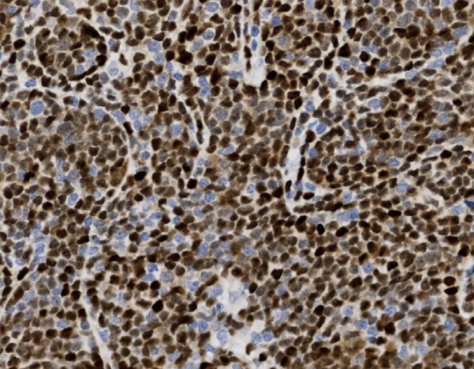
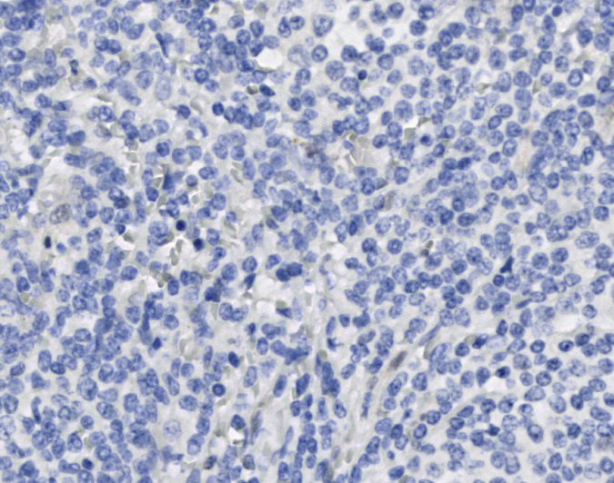
Tissue-Tek Genie® anti-Cytokeratin 5/6
8295-M250: RTU, 250 tests, 1 cartridge; 1 unit
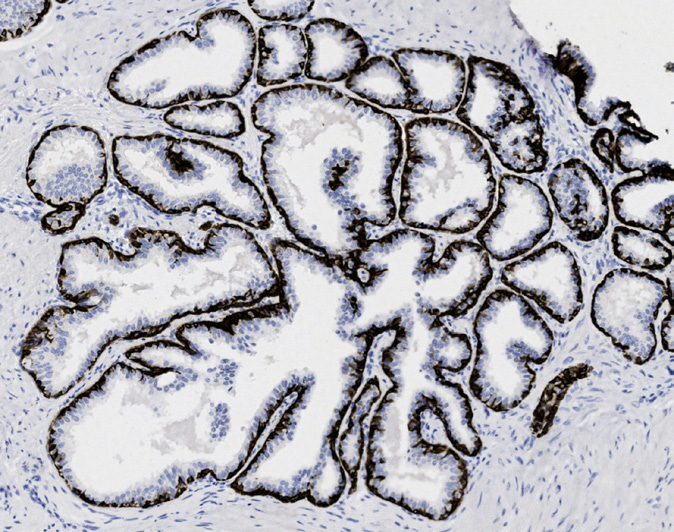
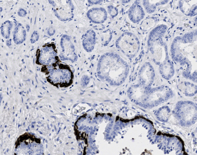
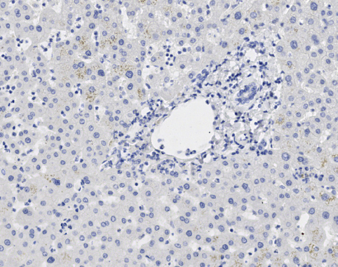
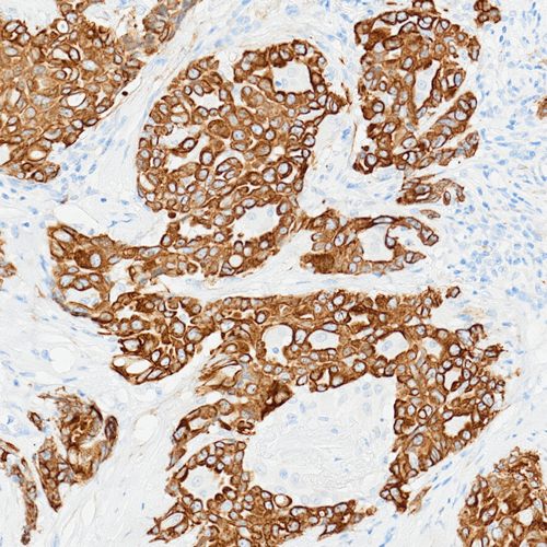
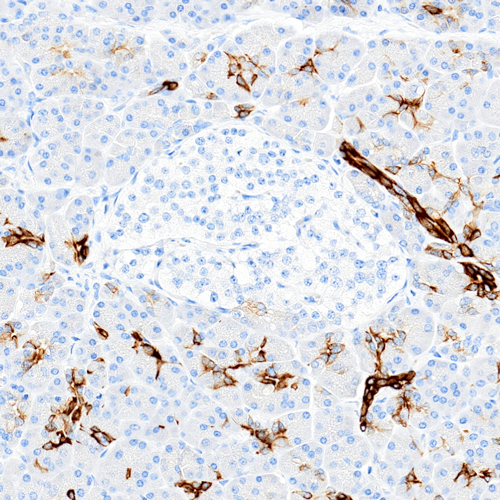
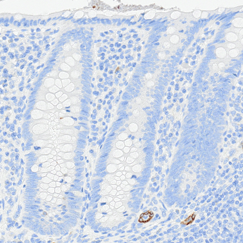
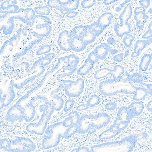
Tissue-Tek Genie® anti-Cytokeratin 7
8296-M250: RTU, 250 tests, 1 cartridge; 1 unit
The Tissue-Tek Genie® anti-Cytokeratin 7 Mouse Monoclonal Antibody [OV-TL 12/30] is designed to qualitatively detect cytokeratin 7 protein in formalin-fixed, paraffin-embedded (FFPE) human tissue specimen sections by immunohistochemistry (IHC) staining on the automated Tissue-Tek Genie® Advanced Staining System.
This device functions as an aid for diagnosis and shall be used by a qualified pathologist with a panel of other antibodies to differentiate adenocarcinomas originating from the lung, breast, endometrium, thyroid gland, or ovary from those arising from the prostate or colorectum.
Tissue-Tek Genie® anti-Cytokeratin 8
8238-M250: RTU, 250 tests, 1 cartridge; 1 unit
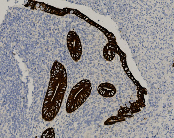
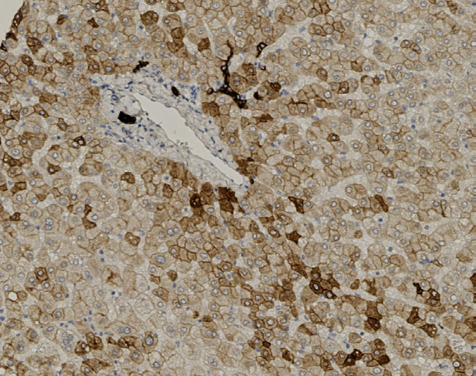
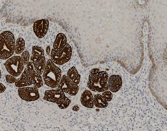
Tissue-Tek Genie® anti-Cytokeratin 14
8300-M250: RTU, 250 tests, 1 cartridge; 1 unit
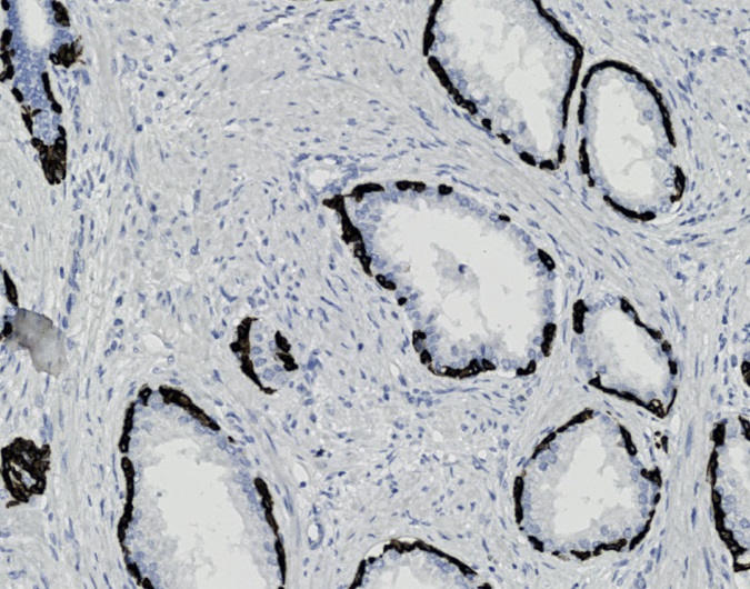
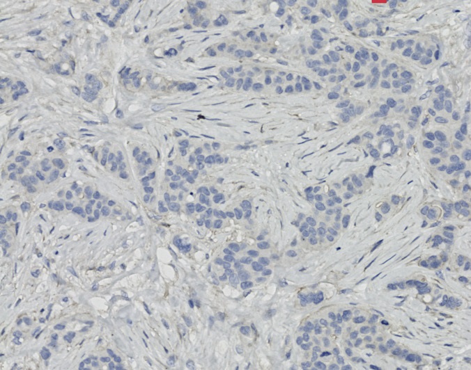
Tissue-Tek Genie® anti-Cytokeratin 18
8302-M250: RTU, 250 tests, 1 cartridge; 1 unit
The Tissue-Tek Genie® anti-Cytokeratin 18 Mouse Monoclonal Antibody [DC10] is designed to qualitatively detect cytokeratin 18 protein in formalin-fixed, paraffin-embedded (FFPE) human tissue specimen sections by immunohistochemistry (IHC) staining on the automated Tissue-Tek Genie® Advanced Staining System.
This device functions as an aid for diagnosis and shall be used by a qualified pathologist with a panel of other antibodies to characterize neoplasms of epithelial origin.
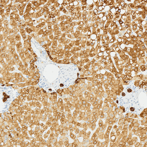
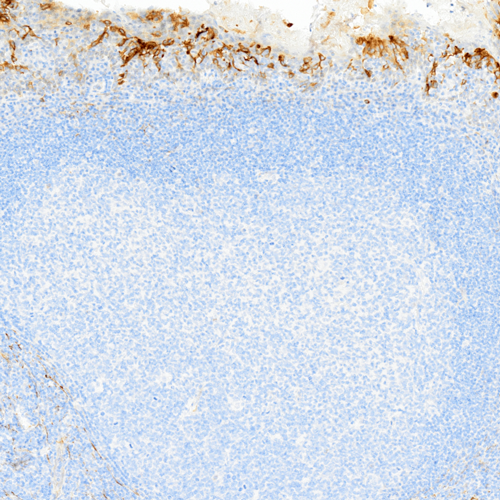
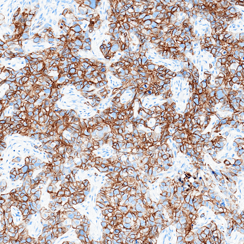

Tissue-Tek Genie® anti-Cytokeratin 19
8303-M250: RTU, 1 cartridge, 250 tests; 1 unit
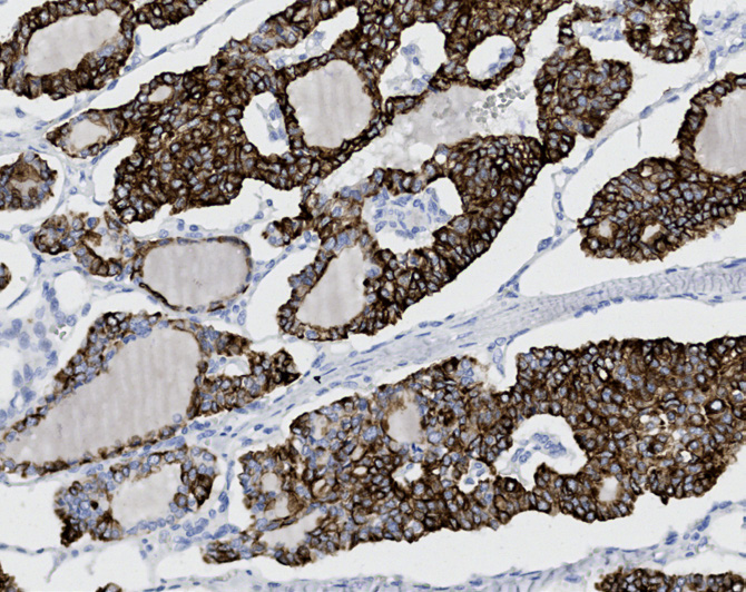
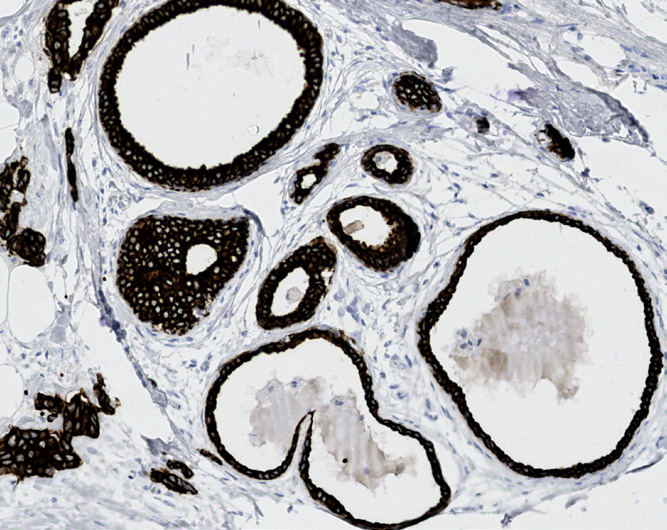
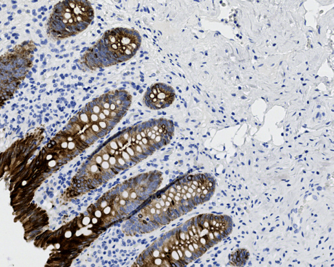
Tissue-Tek Genie® anti-Cytokeratin 20
8304-M250: RTU, 1 cartridge, 250 tests; 1 unit
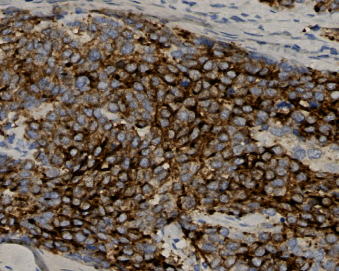
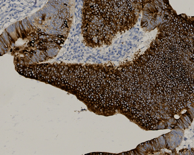
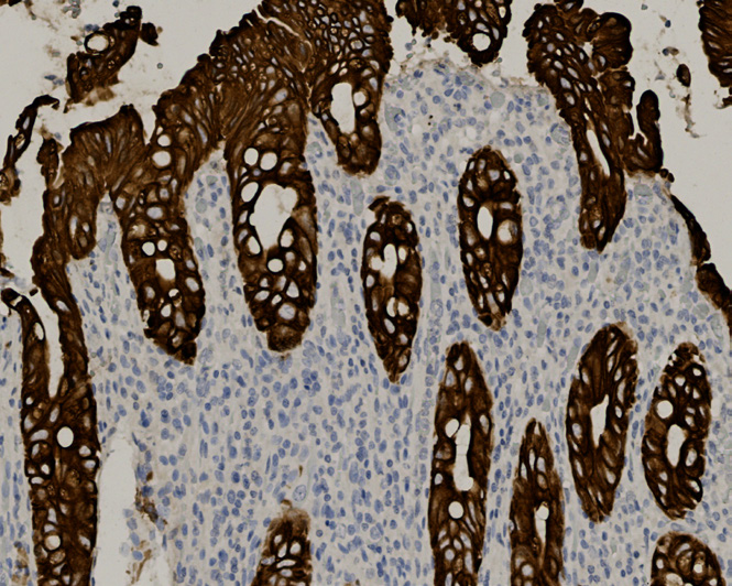
Tissue-Tek Genie® anti-Cytokeratin Antibody Cocktail [AE1/AE3]
8307-M250: RTU, 250 tests, 1 cartridge; 1 unit
![Tissue-Tek Genie® anti-Cytokeratin Antibody Cocktail [AE1/AE3]](https://www.sakuraus.com/wp-content/uploads/2026/02/tissue-tek-geniesupregsupbr-anti-cytokeratin-antibody-cocktail-ae1ae3-product-image.jpg)
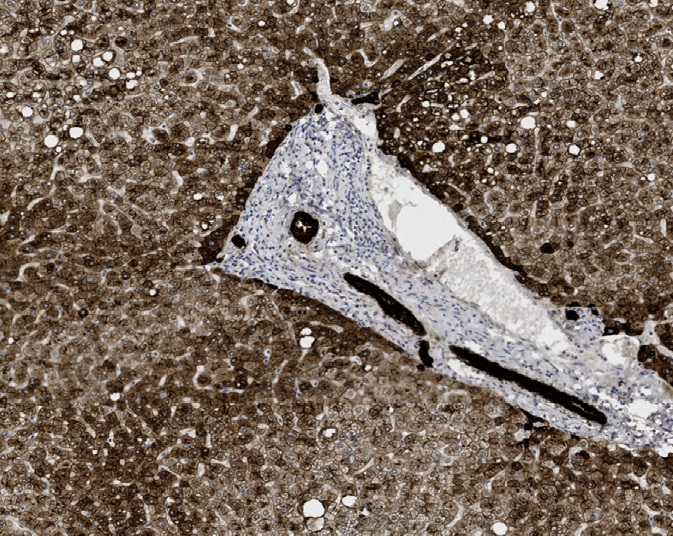
Tissue-Tek Genie® anti-Cytokeratin HMW
8306-M250: RTU, 250 tests, 1 cartridge; 1 unit
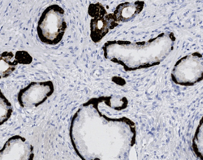
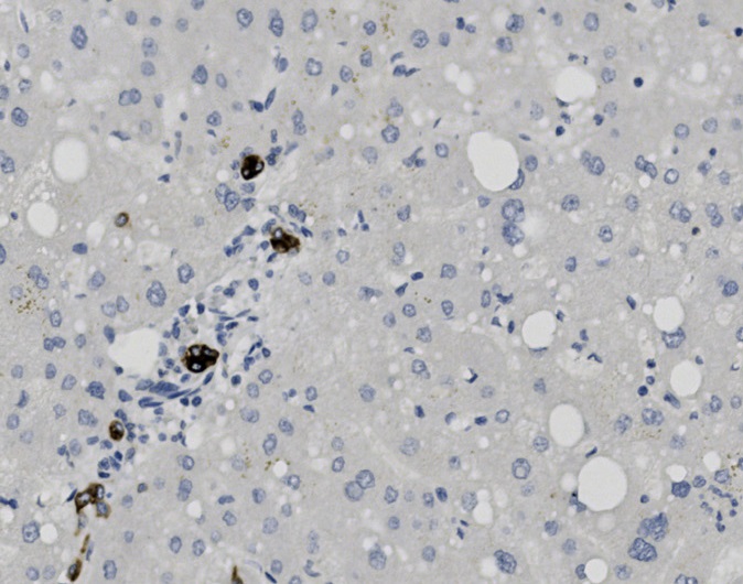
Tissue-Tek Genie® anti-Cytokeratin HMW (CK5)
8294-M250: RTU, 1 cartridge, 250 tests; 1 unit
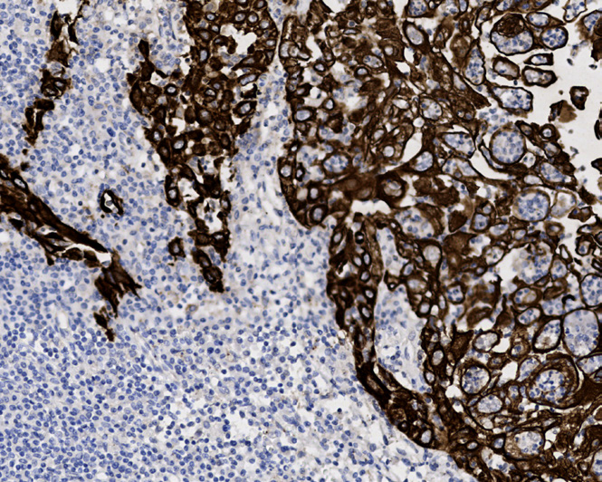
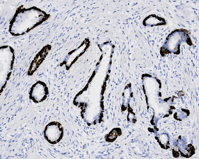
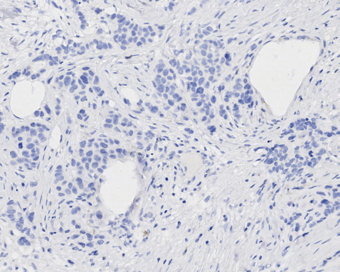
Tissue-Tek Genie® anti-Cytokeratin LMW (CK8/18) Antibody Cocktail [EP17/DC10]
8298-M250: RTU, 250 tests, 1 cartridge; 1 unit
![Tissue-Tek Genie® anti-Cytokeratin LMW (CK8/18) Antibody Cocktail [EP17/DC10]](https://www.sakuraus.com/wp-content/uploads/2026/02/tissue-tek-geniesupregsupbr-anti-cytokeratin-lmw-ck818-antibody-cocktail-ep17dc10-product-image.jpg)
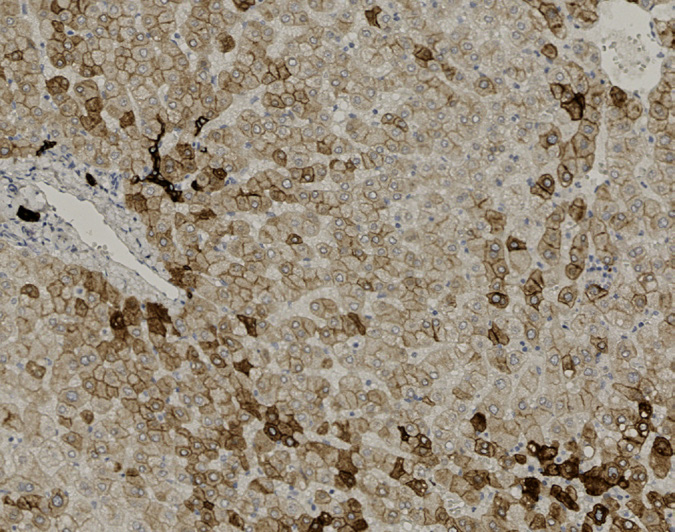
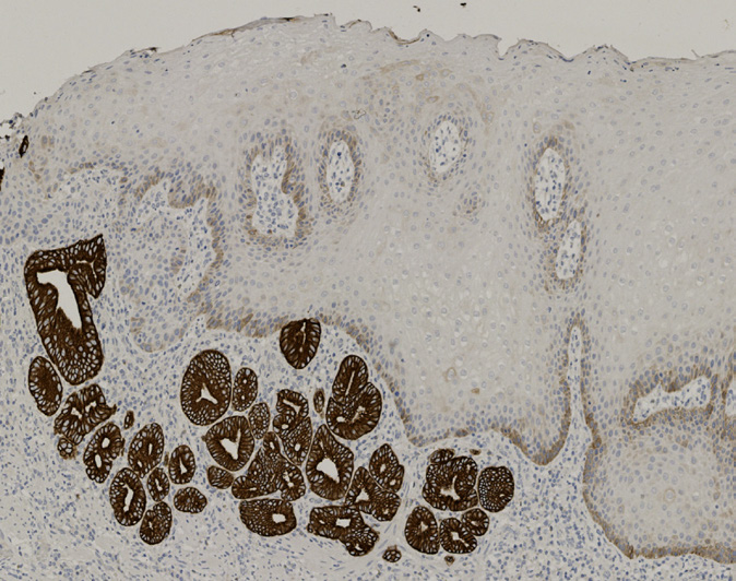
Tissue-Tek Genie® anti-Cytokeratin Pan Antibody Cocktail [AE1/AE3/DC10]
8309-M250: RTU, 250 tests, 1 cartridge; 1 unit
The Tissue-Tek Genie® anti-Cytokeratin Pan Antibody Cocktail [AE1/AE3/DC10] is designed to qualitatively detect cytokeratin protein in formalin-fixed, paraffin-embedded (FFPE) human tissue specimen sections by immunohistochemistry (IHC) staining on the automated Tissue-Tek Genie® Advanced Staining System.
This device functions as an aid for diagnosis and shall be used by a qualified pathologist with a panel of other antibodies to characterize neoplasms of epithelial origin.
![Tissue-Tek Genie® anti-Cytokeratin Pan Antibody Cocktail [AE1/AE3/DC10]](https://www.sakuraus.com/wp-content/uploads/2026/10/pan-ck-3.png)
![Tissue-Tek Genie® anti-Cytokeratin Pan Antibody Cocktail [AE1/AE3/DC10] - Additional Image 1](https://www.sakuraus.com/wp-content/uploads/2026/10/pan-ck-1.png)
![Tissue-Tek Genie® anti-Cytokeratin Pan Antibody Cocktail [AE1/AE3/DC10] - Additional Image 2](https://www.sakuraus.com/wp-content/uploads/2026/10/pan-ck-2.png)
![Tissue-Tek Genie® anti-Cytokeratin Pan Antibody Cocktail [AE1/AE3/DC10] - Additional Image 3](https://www.sakuraus.com/wp-content/uploads/2026/10/pan-ck-4.png)
Tissue-Tek Genie® anti-Desmin
8311-M250: RTU, 1 cartridge, 250 tests; 1 unit
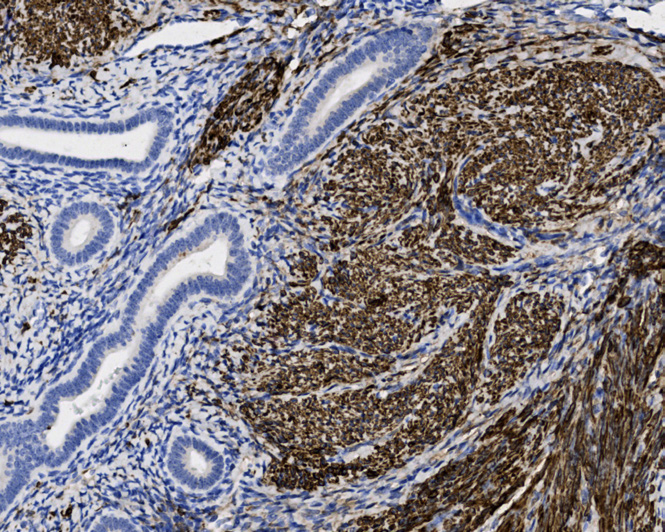
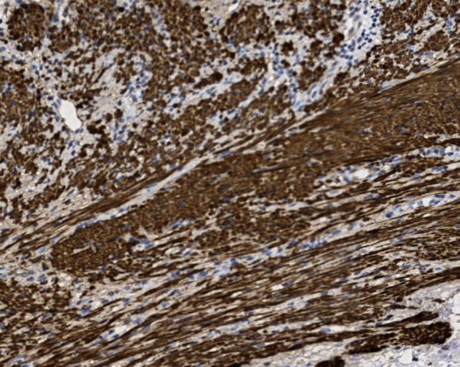
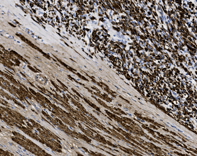
Tissue-Tek Genie® anti-DOG1
8368-M250: RTU, 1 cartridge, 250 tests; 1 unit
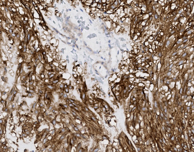
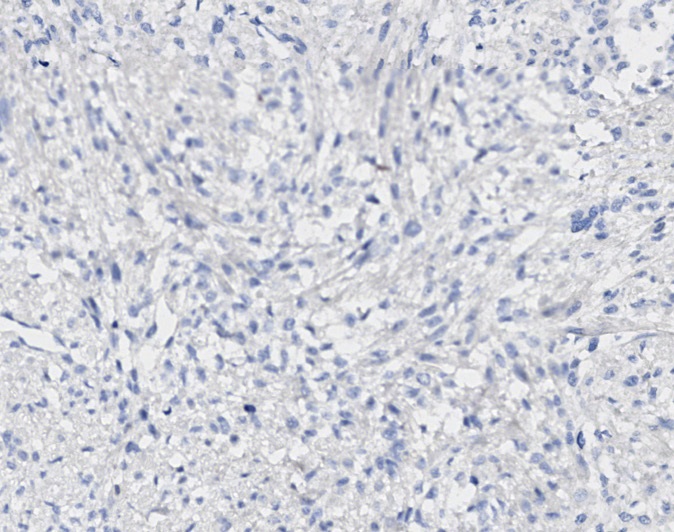
Tissue-Tek Genie® anti-E-Cadherin
8269-M250: RTU, 250 tests, 1 cartridge; 1 unit
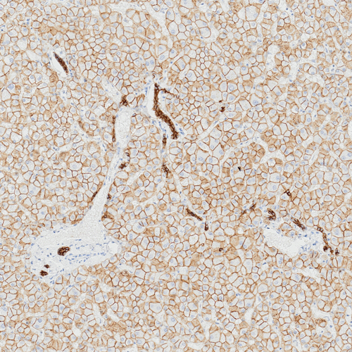
The Tissue-Tek Genie® anti-E-Cadherin Mouse Monoclonal Antibody [ZM63] is designed to qualitatively detect E-cadherin protein in formalin-fixed, paraffin-embedded (FFPE) human tissue specimen sections by immunohistochemistry (IHC) staining on the automated Tissue-Tek Genie® Advanced Staining System.
This device functions as an aid for diagnosis and shall be used by a qualified pathologist with a panel of other antibodies to differentiate ductal from lobular breast carcinomas.
Tissue-Tek Genie® anti-Epithelial Cell Adhesion Molecule [BS14]
8337-M250: RTU, 250 tests, 1 cartridge; 1 unit
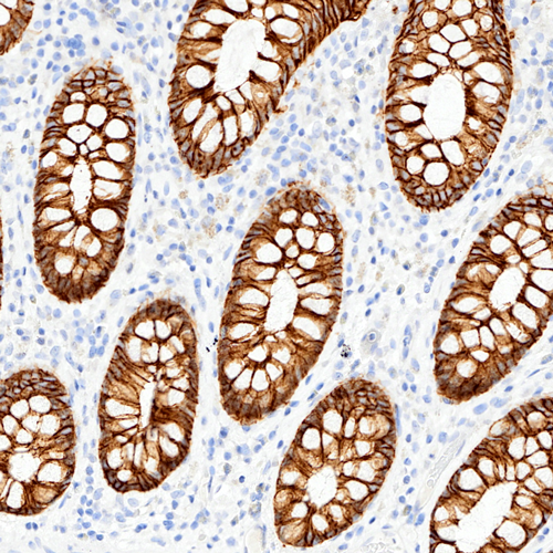
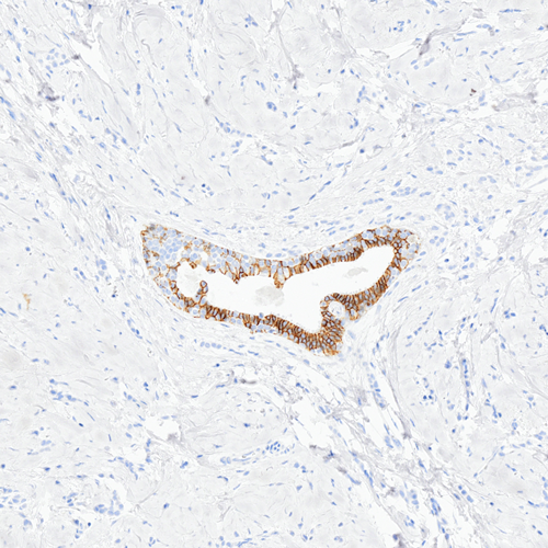
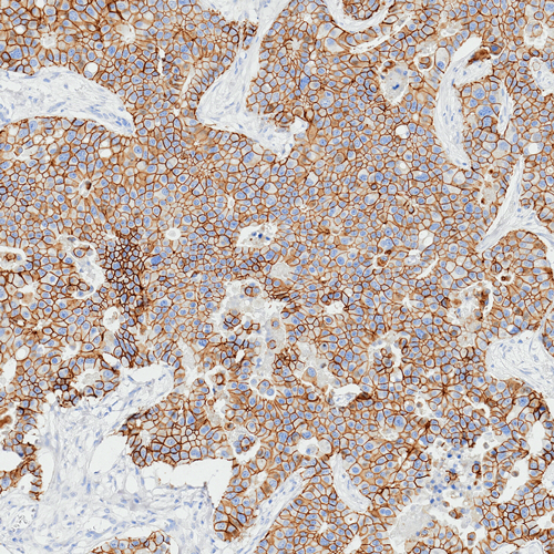
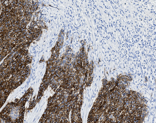
The Tissue-Tek Genie® anti-Epithelial Cell Adhesion Molecule Mouse Monoclonal Antibody [BS14] is designed to qualitatively detect EpCAM protein in formalin-fixed, paraffin-embedded (FFPE) human tissue specimen sections by immunohistochemistry (IHC) staining on the automated Tissue-Tek Genie® Advanced Staining System. This device functions as an aid for diagnosis and shall be used by a qualified pathologist with a panel of other antibodies to differentiate lung adenocarcinoma from mesothelioma and basal cell carcinoma from squamous cell carcinoma. EpCAM expression is commonly found in the majority of adenocarcinomas. However, it is often used to differentiate lung adenocarcinoma from mesotheliomas. However, the lack of EpCAM immunoreactivity has been used to aid in distinguishing pleural mesothelioma from lung adenocarcinomas. In recent studies, EpCAM has also been used to differentiate basal cell carcinomas from squamous cell carcinomas of ectodermal origin.
![Tissue-Tek Genie® anti-Epithelial Cell Adhesion Molecule [BS14]](https://www.sakuraus.com/wp-content/uploads/2026/09/tissue-tek-geniereg-anti-epithelial-cell-adhesion-molecule-bs14-product-image.jpg)
![Tissue-Tek Genie<sup>®</sup> anti-Epithelial Cell Adhesion Molecule [BS14]](https://www.sakuraus.com/wp-content/uploads/2026/09/tissue-tek-geniereg-anti-epithelial-cell-adhesion-molecule-bs14-additional-image-1.jpg)
![Tissue-Tek Genie<sup>®</sup> anti-Epithelial Cell Adhesion Molecule [BS14]](https://www.sakuraus.com/wp-content/uploads/2026/09/tissue-tek-geniereg-anti-epithelial-cell-adhesion-molecule-bs14-additional-image-2.jpg)
![Tissue-Tek Genie<sup>®</sup> anti-Epithelial Cell Adhesion Molecule [BS14]](https://www.sakuraus.com/wp-content/uploads/2026/09/tissue-tek-geniereg-anti-epithelial-cell-adhesion-molecule-bs14-additional-image-3.jpg)
Tissue-Tek Genie® anti-Epithelial Cell Adhesion Molecule [VU-1D9]
8230-M250: RTU, 1 cartridge, 250 tests; 1 unit
![Tissue-Tek Genie® anti-Epithelial Cell Adhesion Molecule [VU-1D9]](https://www.sakuraus.com/wp-content/uploads/2026/02/tissue-tek-geniesupregsupbr-anti-epithelial-cell-adhesion-molecule-product-image.jpg)
Tissue-Tek Genie® anti-Epithelial Membrane Antigen
8233-M250: RTU, 1 cartridge, 250 tests; 1 unit
Tissue-Tek Genie® anti-ERG
8323-M250: RTU, 250 tests, 1 cartridge; 1 unit
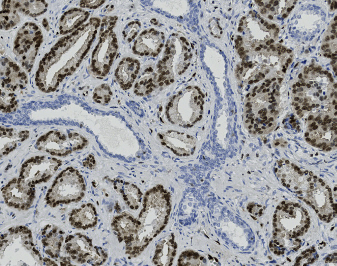
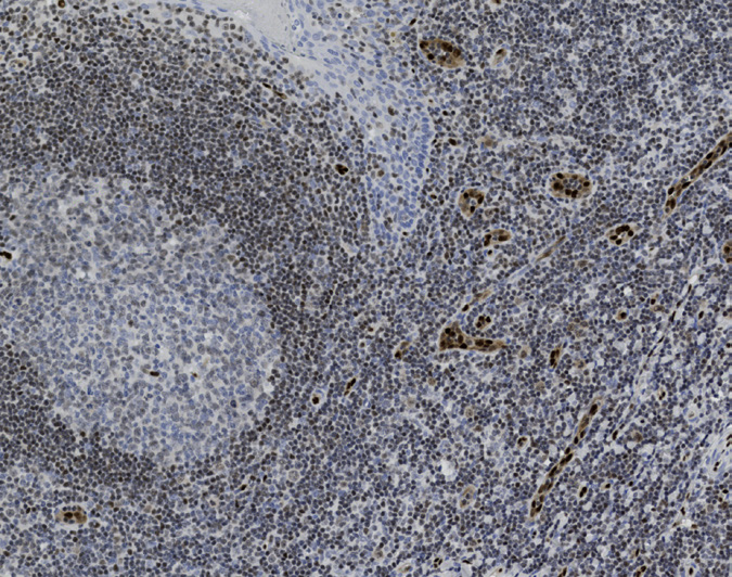
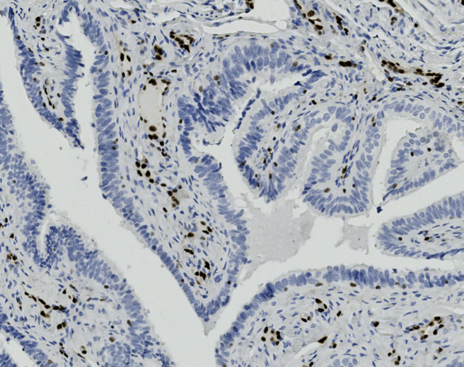
45-65% of prostate cancers. ERG is expressed in endothelial neoplasms including hemangioendothelioma, angiosarcoma and Kaposi sarcoma. Clone EP111 is also named as EPR3864. This clone is capable of detecting both wild-type and truncated ERG.
Tissue-Tek Genie® anti-Factor XIIIa
8293-M250: RTU, 1 cartridge, 250 tests; 1 unit
Tissue-Tek Genie® anti-Gastrin
8314-M250: RTU, 1 cartridge, 250 tests; 1 unit
Tissue-Tek Genie® anti-GATA3
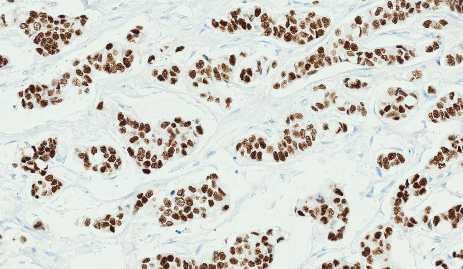
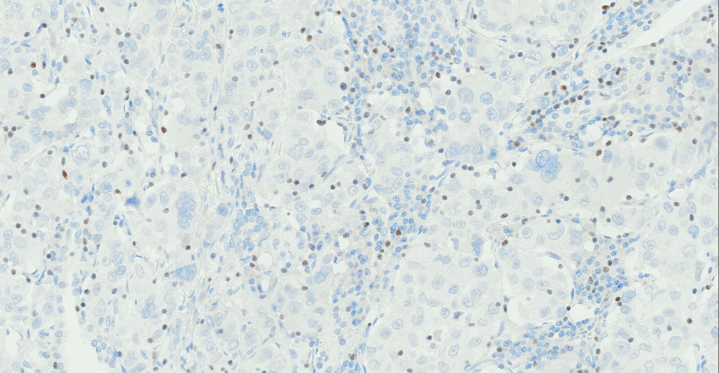
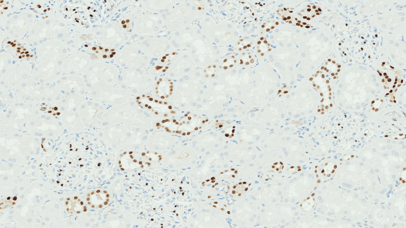
8357-M250: RTU, 250 tests, 1 cartridge; 1
GATA binding protein 3 (GATA3) is a transcription factor that plays a crucial role in the development and differentiation of various tissues.
Tissue-Tek Genie® anti-GCDFP-15
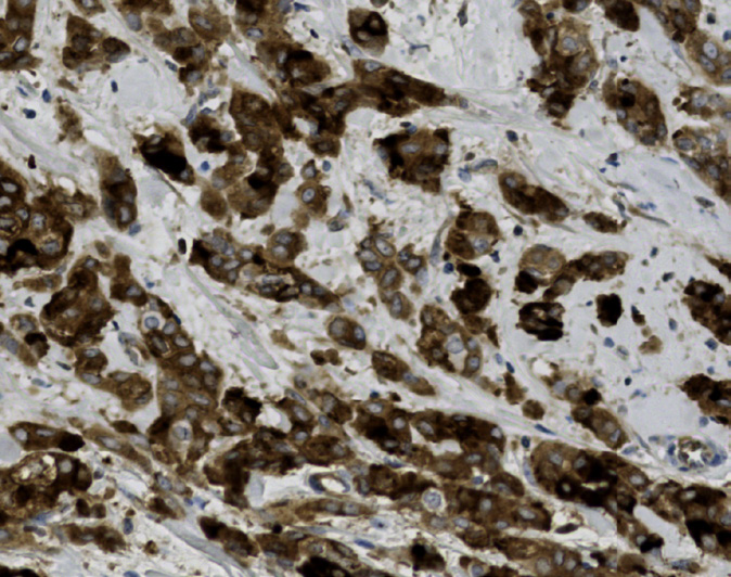
8325-M250: RTU, 250 tests, 1 cartridge; 1 unit
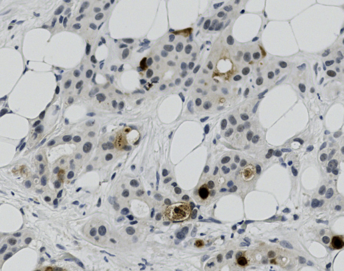
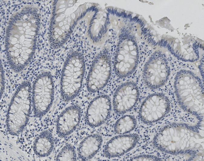
Gross cystic disease fluid protein-15 (GCDFP-15) is a secretory glycoprotein and is expressed in the acinic cells of the breast, salivary glands, sweat glands, and seminal vesicle. GCDFP-15 is often expressed in neoplastic cells of breast carcinomas (50-70%), mammary and extramammary Paget’s disease, sweat gland tumors (particularly apocrine), and salivary gland tumors.
Tissue-Tek Genie® anti-Glial Fibrillary Acidic Protein (GFAP)
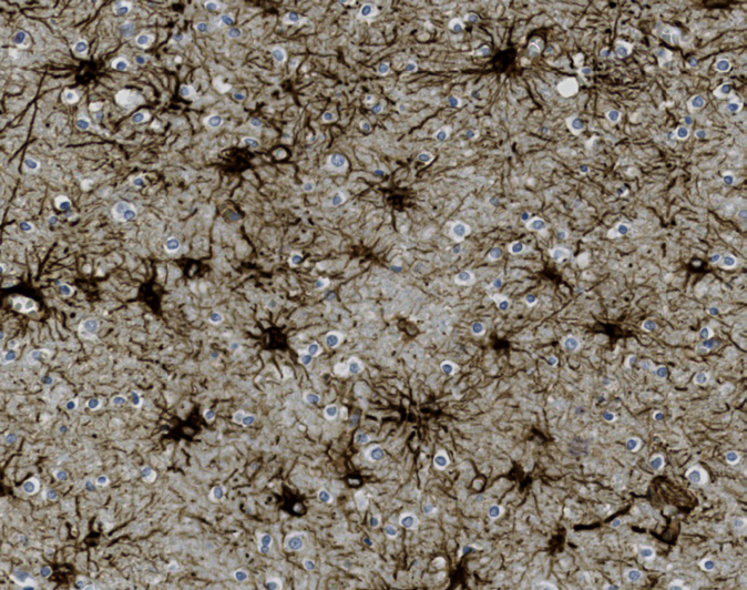
8316-M250: RTU, 250 tests, 1 cartridge; 1 unit
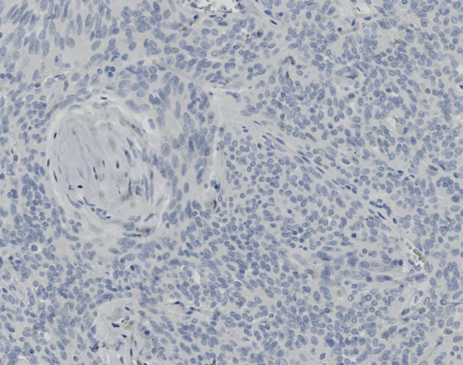
Tissue-Tek Genie® anti-Glypican-3
8346-M250: RTU, 250 tests, 1 cartridge; 1 unit
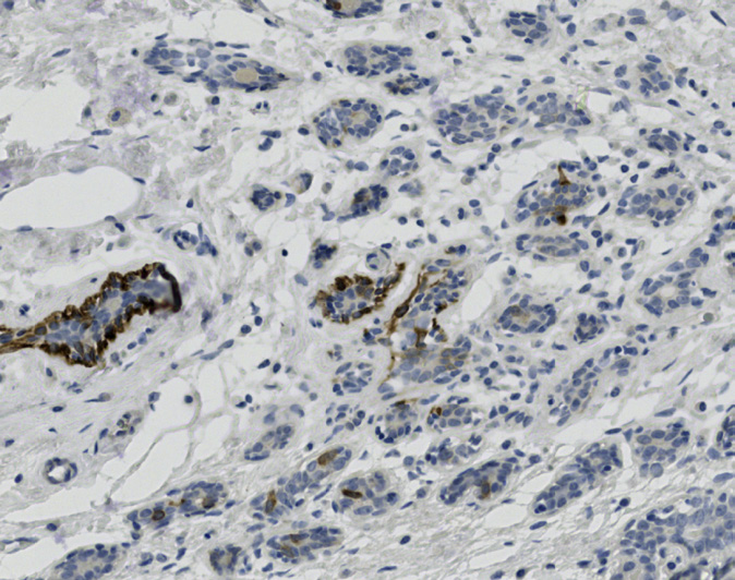
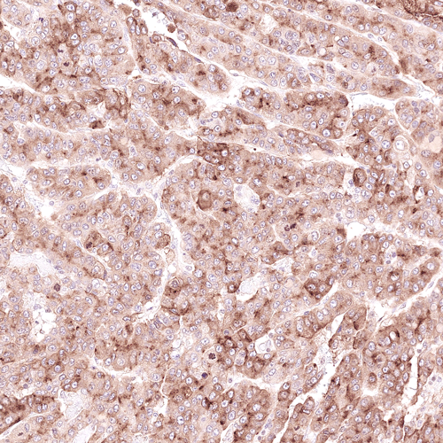
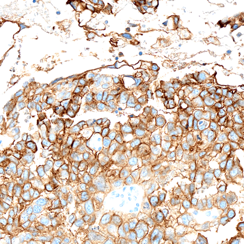
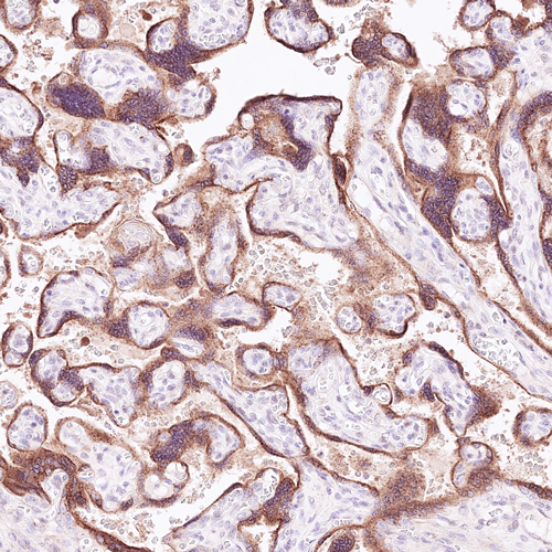
The Tissue-Tek Genie® anti-Glypican-3 Mouse Monoclonal Antibody [1G12] is designed to qualitatively detect Glypican-3 protein in formalin-fixed, paraffin-embedded (FFPE) human tissue specimen sections by immunohistochemistry (IHC) staining on the automated Tissue-Tek Genie® Advanced Staining System.
This device functions as an aid for diagnosis and shall be used by a qualified pathologist with a panel of other antibodies to identify hepatocellular carcinoma and yolk sac tumors.
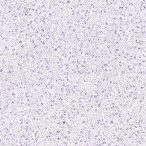
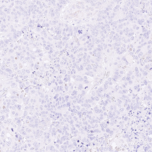
Tissue-Tek Genie® anti-Granzyme B
8234-M250: RTU, 250 tests, 1 cartridge; 1 unit
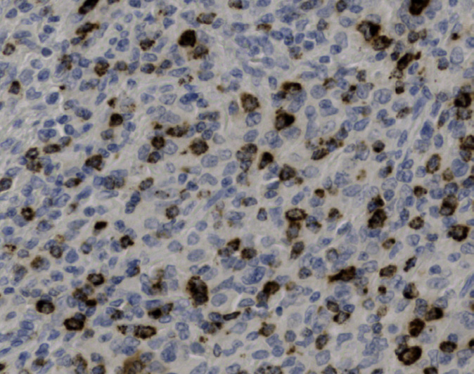
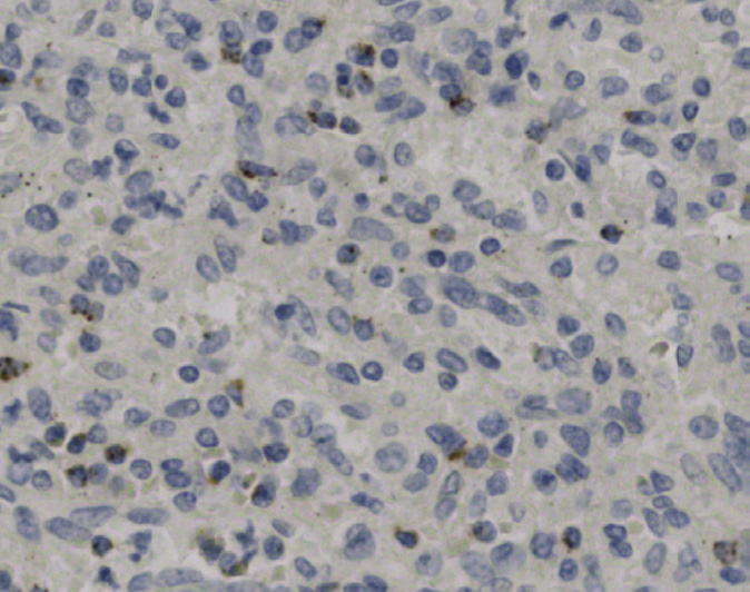
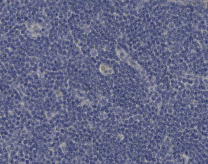
Tissue-Tek Genie® anti-Helicobacter pylori
8317-M250: RTU, 1 cartridge, 250 tests; 1 unit
Tissue-Tek Genie® anti-Hepatocyte Specific Antigen (HEP PAR 1)
8374-M250: RTU, 250 tests, 1 cartridge; 1 unit
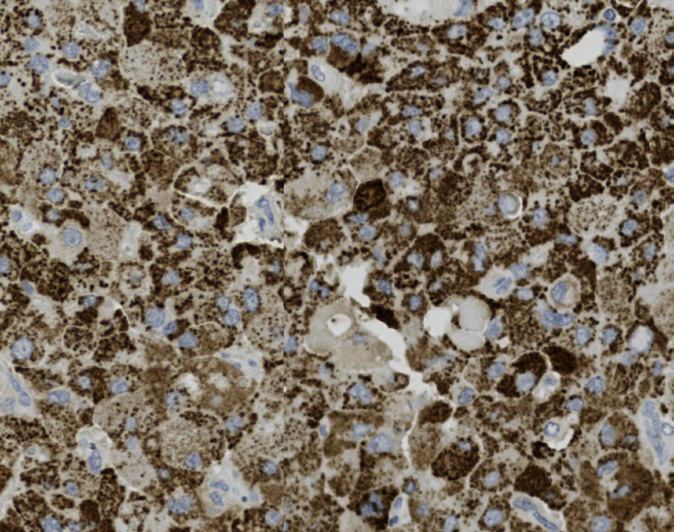
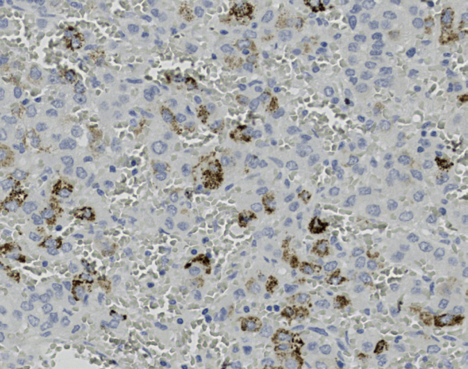
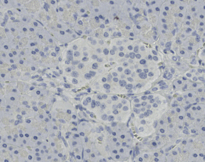
Tissue-Tek Genie® anti-HHV8
8378-M250: RTU, 1 cartridge, 250 tests; 1 unit
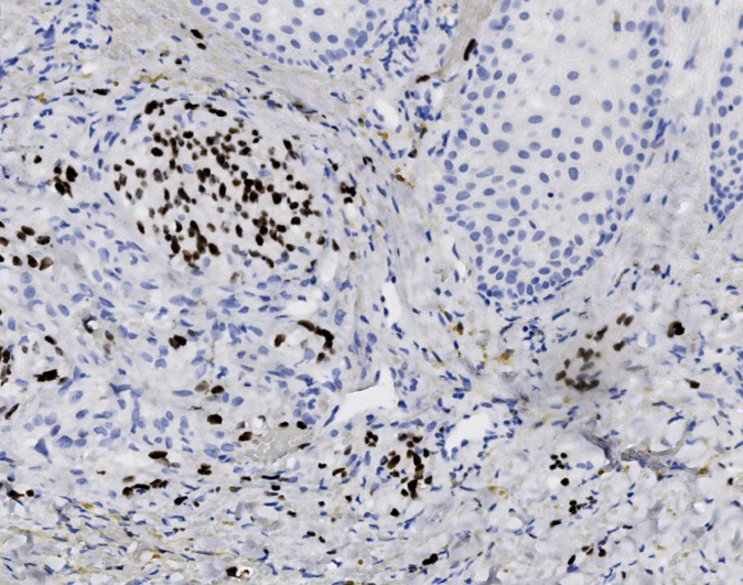
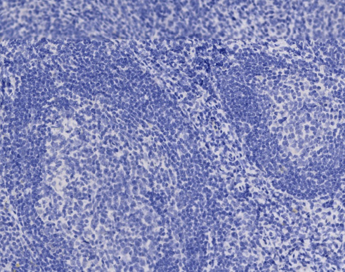
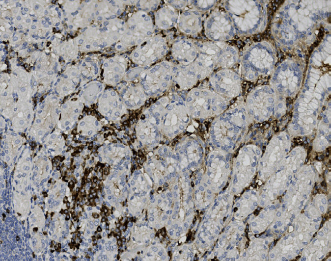
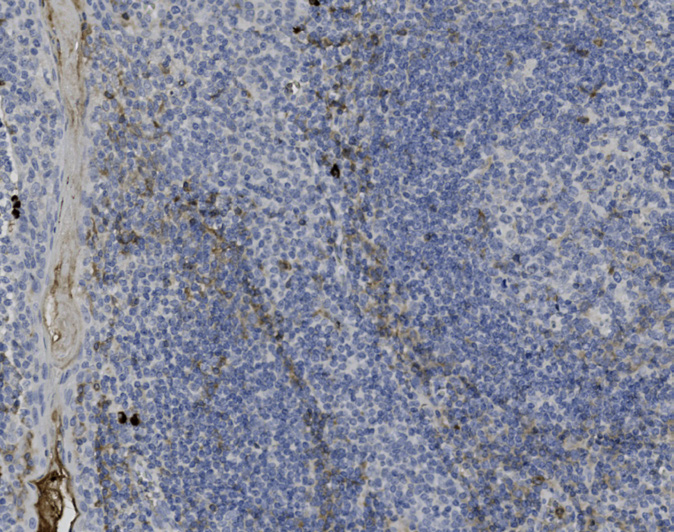
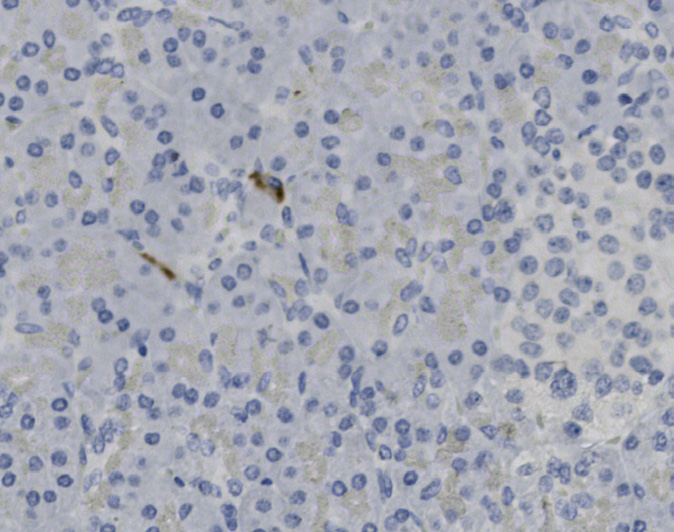
Tissue-Tek Genie® anti-IgA
8229-M250: RTU, 250 tests, 1 cartridge; 1 unit
Tissue-Tek Genie® anti-IgD
8382-M250: RTU, 250 tests, 1 cartridge; 1 unit
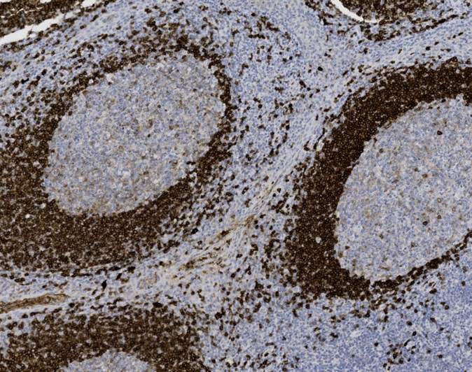
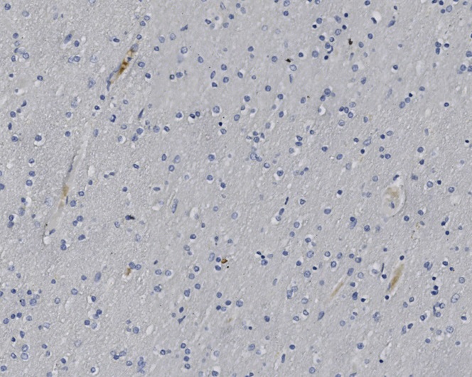
Tissue-Tek Genie® anti-IgG
8383-M250: RTU, 250 tests, 1 cartridge; 1 unit
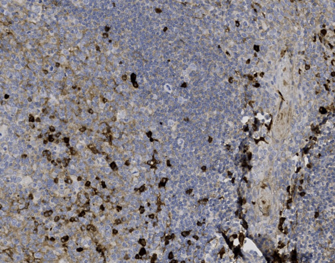
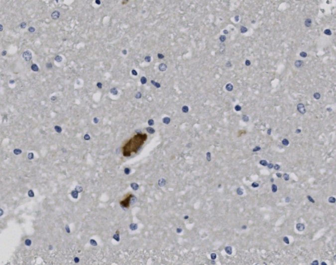
Tissue-Tek Genie® anti-IgG4
8384-M250: RTU, 250 tests, 1 cartridge; 1 unit
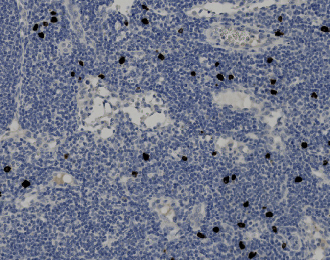
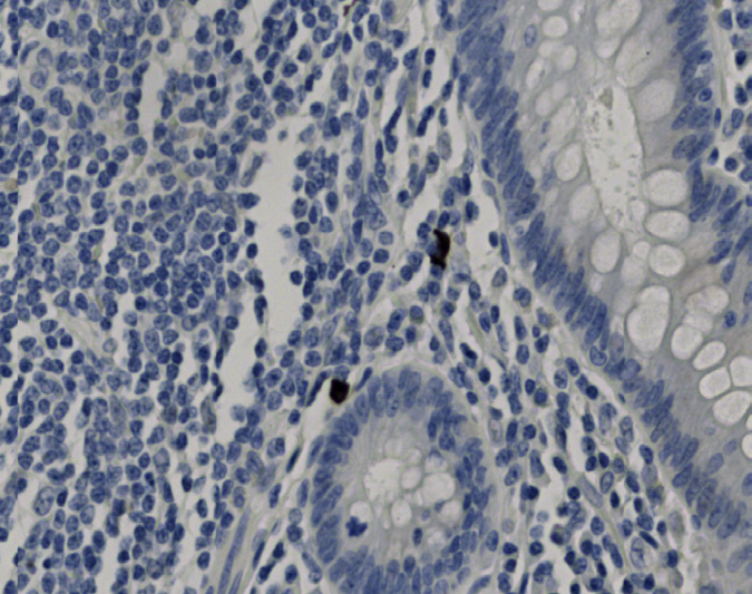
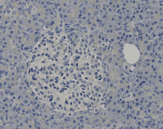
Tissue-Tek Genie® anti-IgM
8385-M250: RTU, 250 tests, 1 cartridge; 1 unit
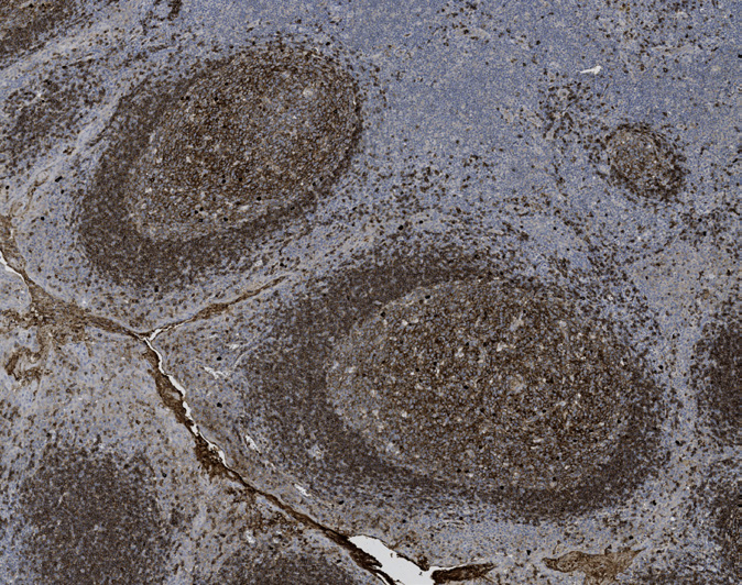
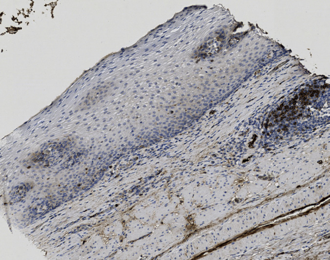
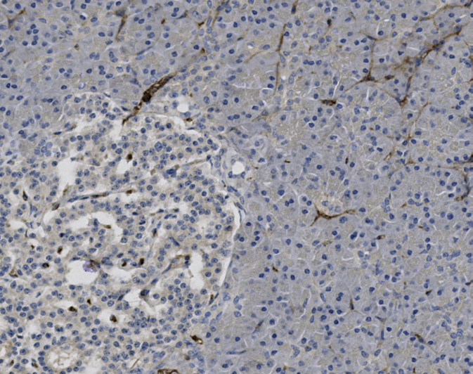
Tissue-Tek Genie® anti-INSM1
8379-M250: RTU, 250 tests, 1 cartridge; 1 unit
The Tissue-Tek Genie® anti-INSM1 Rabbit Monoclonal Antibody [ZR395] is designed to qualitatively detect INSM1 protein in formalin-fixed, paraffin-embedded (FFPE) human tissue specimen sections by immunohistochemistry (IHC) staining on the automated Tissue-Tek Genie® Advanced Staining System.
This device functions as an aid for diagnosis and shall be used by a qualified pathologist with a panel of other antibodies to identify neuroendocrine neoplasms. Recent studies suggest that INSM1 is a highly specific biomarker for neuroendocrine differentiation, demonstrating greater specificity than chromogranin A (CGA) and comparable or superior sensitivity to synaptophysin (SYP) and CD56. INSM1 is detected in neoplastic cells of neuroendocrine neoplasms, including small cell lung carcinoma and medullary thyroid carcinoma.
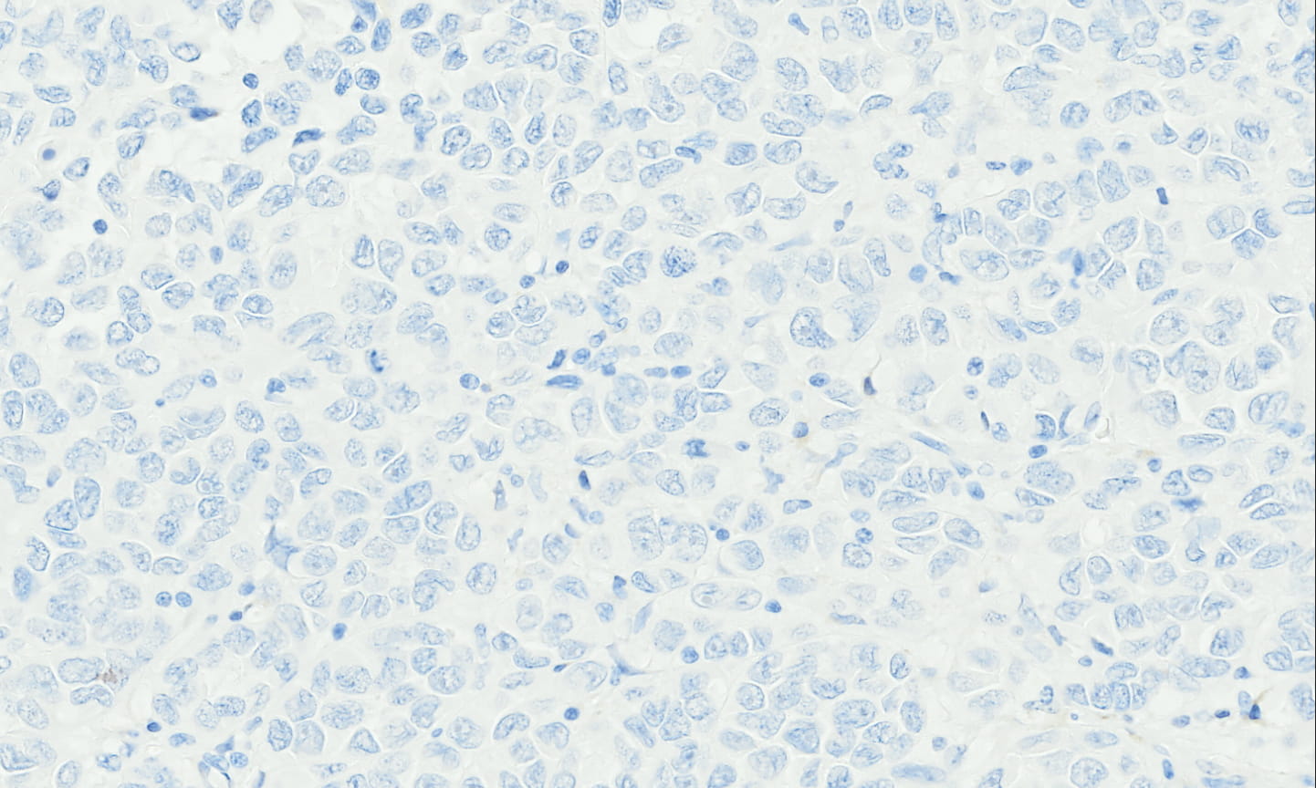
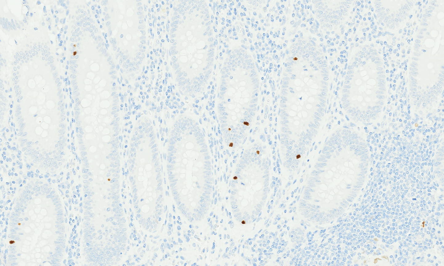
Tissue-Tek Genie® anti-Kappa Light Chains
8394-M250: RTU, 250 tests, 1 cartridge; 1 unit
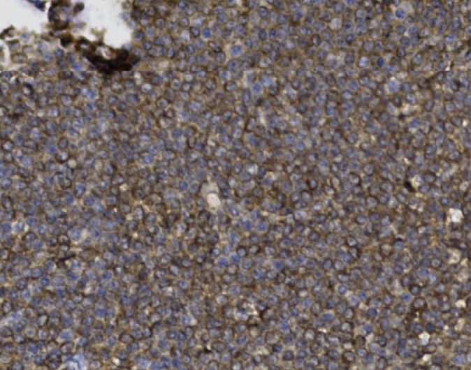
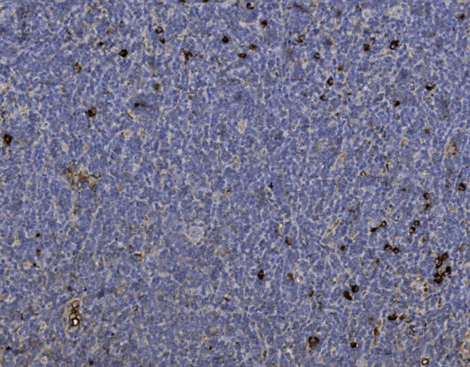
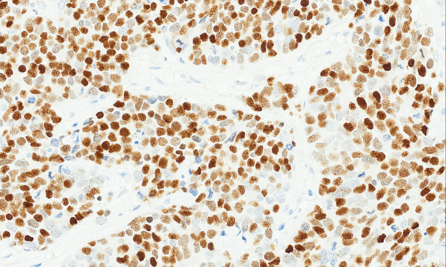
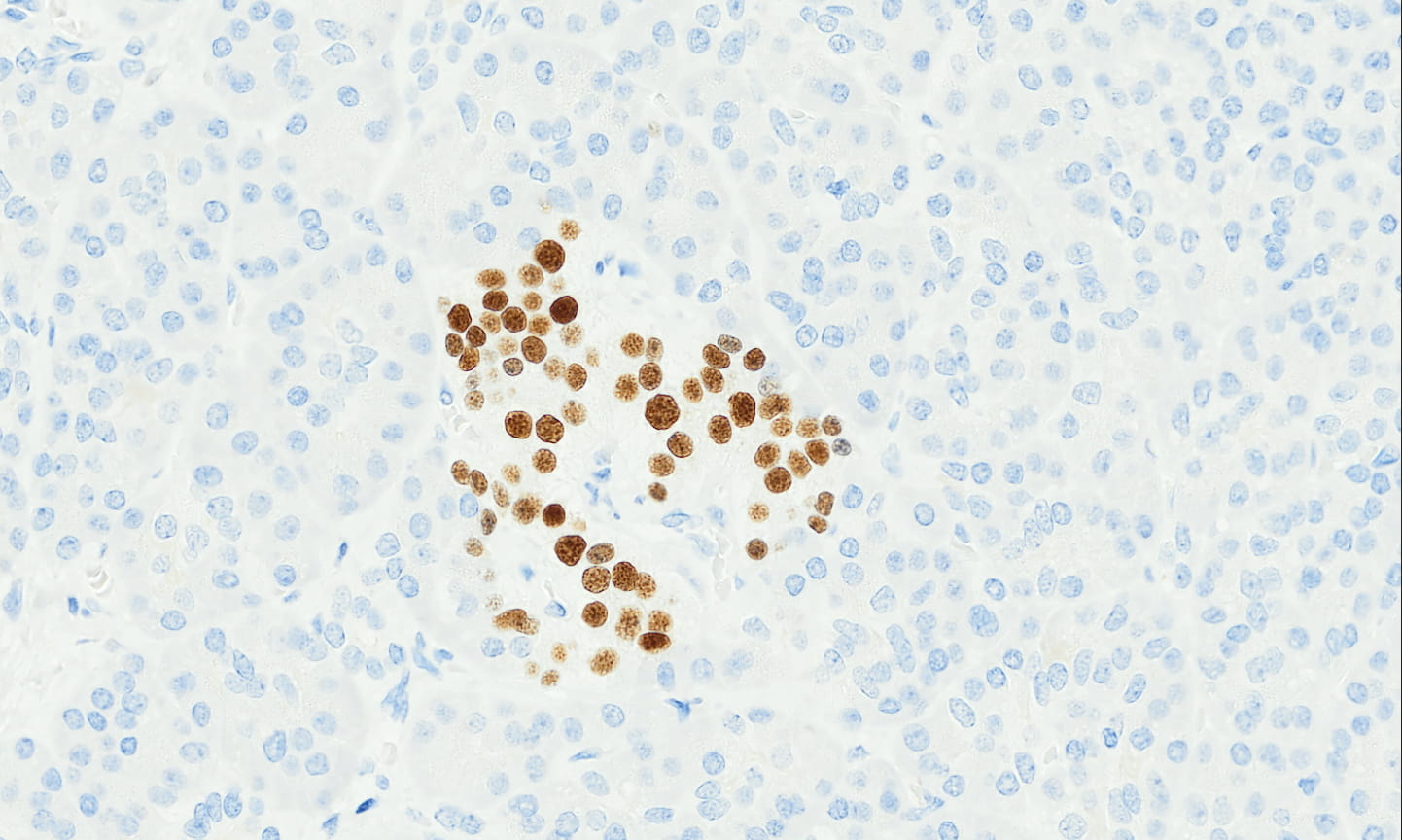
Tissue-Tek Genie® anti-Ki67
8473-M250 1 cartridge, 250 tests, RTU; 1 unit
Ki67 is a proliferation-associated nuclear protein expressed during all active phases of the cell cycle and is absent in resting cells.
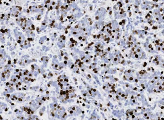
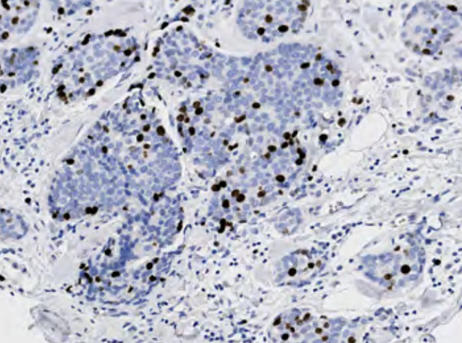
Tissue-Tek Genie® anti-Ki67 [GM027]
8276-M250: RTU, 250 tests, 1 cartridge; 1 unit
The Tissue-Tek Genie® anti-Ki67 Mouse Monoclonal Antibody [GM027] is designed to qualitatively detect Ki67 protein in formalin-fixed, paraffin-embedded (FFPE) human tissue specimen sections by immunohistochemistry (IHC) staining on the automated Tissue-Tek Genie® Advanced Staining System.
This device functions as an aid for diagnosis and shall be used by a qualified pathologist with a panel of other antibodies to assess the proliferative index of various tumors.
![Tissue-Tek Genie® anti-Ki67 [GM027]](https://www.sakuraus.com/wp-content/uploads/2026/10/ki67-gm027-1.png)
![Tissue-Tek Genie® anti-Ki67 [GM027] - Additional Image 1](https://www.sakuraus.com/wp-content/uploads/2026/10/ki67-gm027-2.png)
![Tissue-Tek Genie® anti-Ki67 [GM027] - Additional Image 2](https://www.sakuraus.com/wp-content/uploads/2026/10/ki67-gm027-3.png)
![Tissue-Tek Genie® anti-Ki67 [GM027] - Additional Image 3](https://www.sakuraus.com/wp-content/uploads/2026/10/ki67-gm027-4.png)
Tissue-Tek Genie® anti-Lambda Light Chains
8398-M250: RTU, 250 tests, 1 cartridge; 1 unit
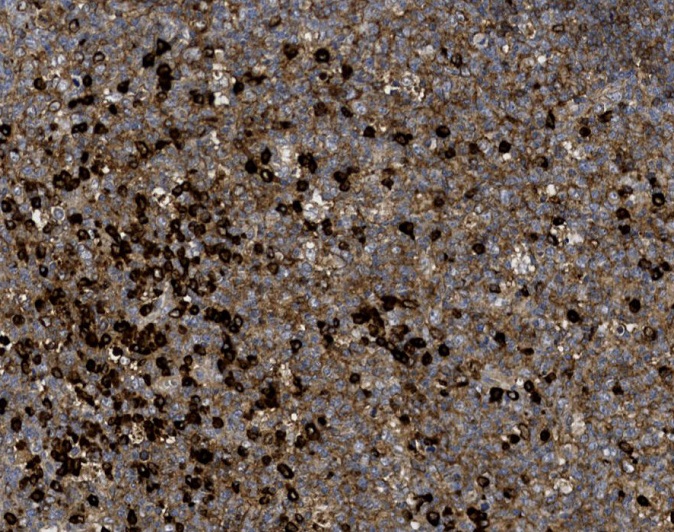
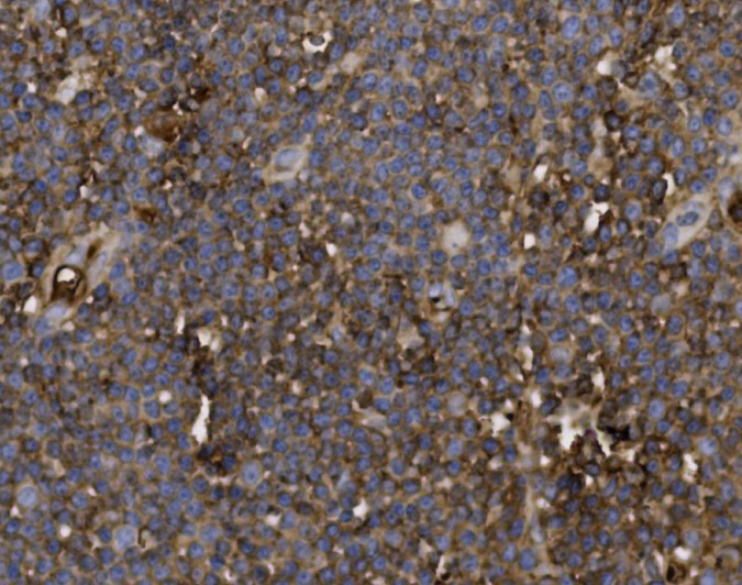
Tissue-Tek Genie® anti-Lysozyme
8333-M250: RTU, 250 tests, 1 cartridge; 1 unit
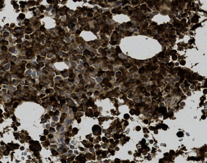
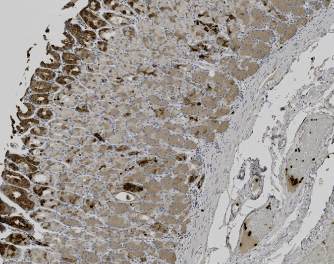
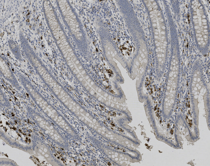
Tissue-Tek Genie® anti-Mammaglobin
8240-M250: RTU, 250 tests, 1 cartridge; 1 unit
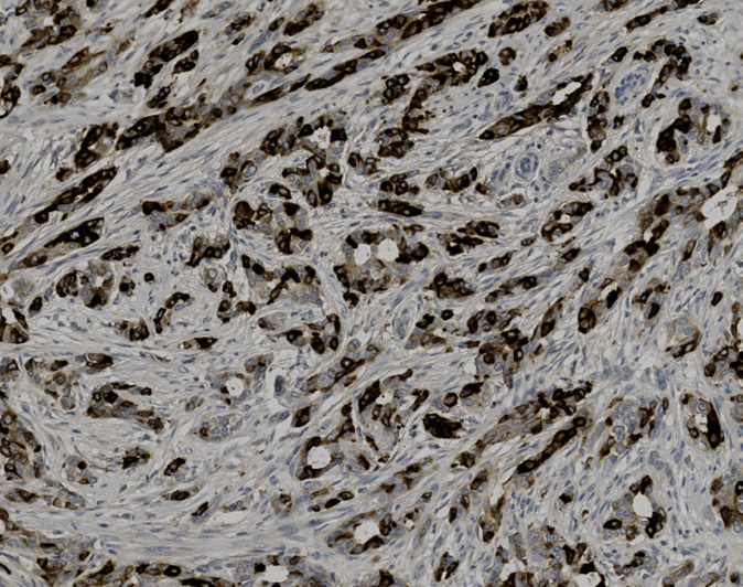
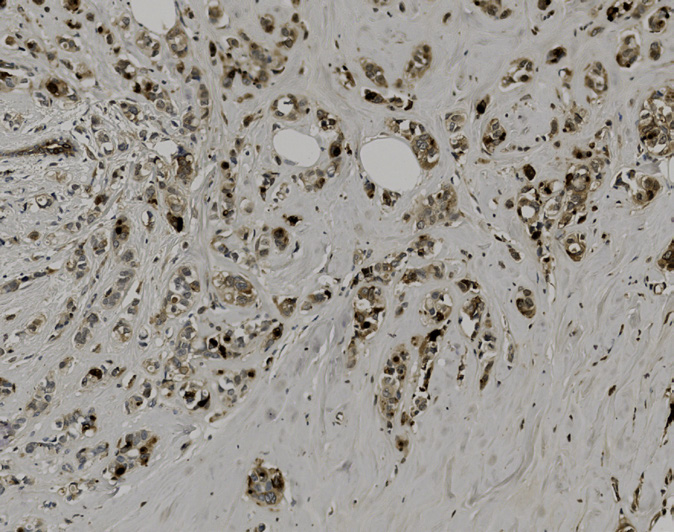
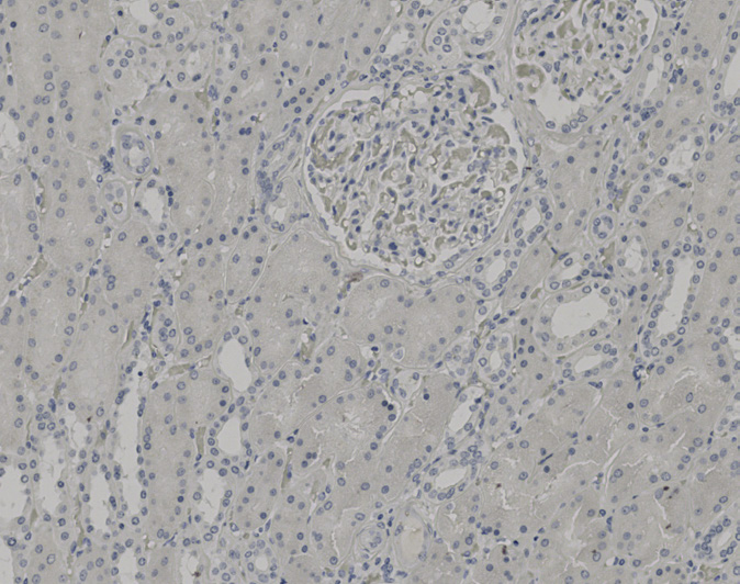
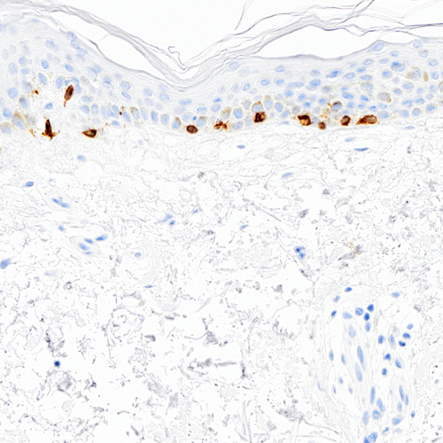
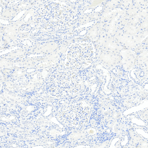
Tissue-Tek Genie® anti-Melan-A
8319-M250: RTU, 250 tests, 1 cartridge; 1 unit
The Tissue-Tek Genie® anti-Melan A Rabbit Monoclonal Antibody [EP43] is designed to qualitatively detect Melan A protein in formalin-fixed, paraffin-embedded (FFPE) human tissue specimen sections by immunohistochemistry (IHC) staining on the automated Tissue-Tek Genie® Advanced Staining System.
This device functions as an aid for diagnosis and shall be used by a qualified pathologist with a panel of other antibodies to identify melanomas.
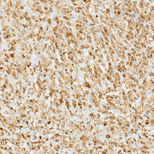
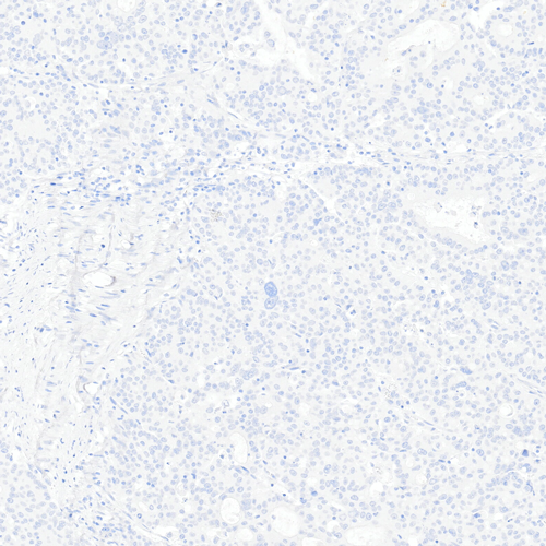
Tissue-Tek Genie® anti-Melanoma Pan Antibody Cocktail [HMB45/EP43/T311]
8371-M250: RTU, 250 tests, 1 cartridge; 1 unit
![Tissue-Tek Genie® anti-Melanoma Pan Antibody Cocktail [HMB45/EP43/T311]](https://www.sakuraus.com/wp-content/uploads/2026/02/tissue-tek-geniesupregsupbr-anti-melanoma-pan-antibody-cocktail-hmb45ep43t311-product-image.jpg)
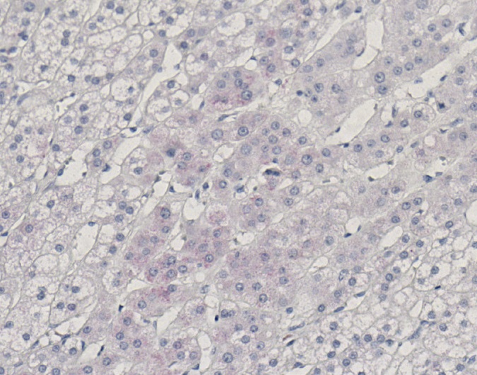
Tissue-Tek Genie® anti-Melanosome
8232-M250: RTU, 1 cartridge, 250 tests; 1 unit
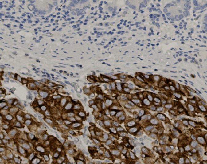
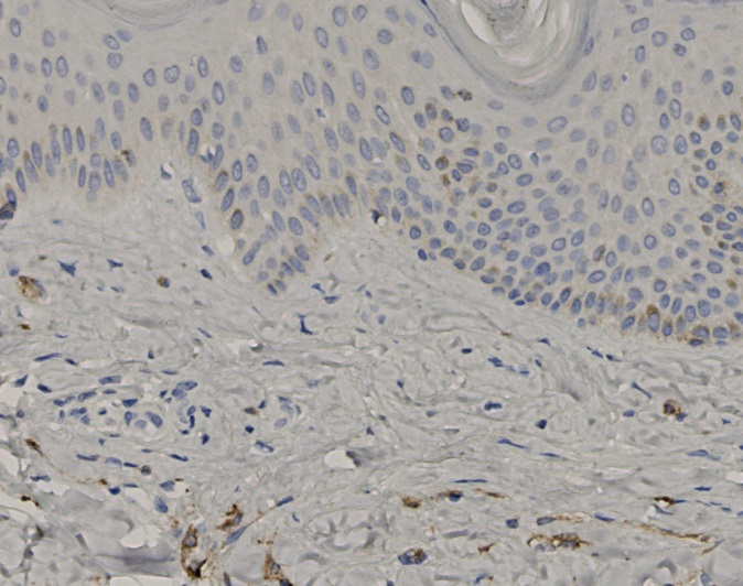
Tissue-Tek Genie® anti-MLH1
8324-M250: RTU, 1 cartridge, 250 tests; 1 unit
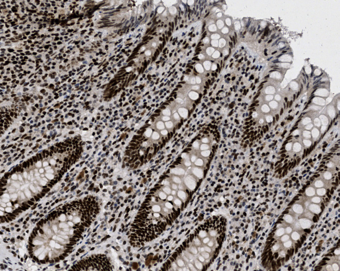
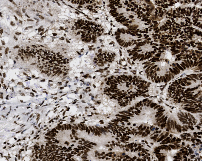
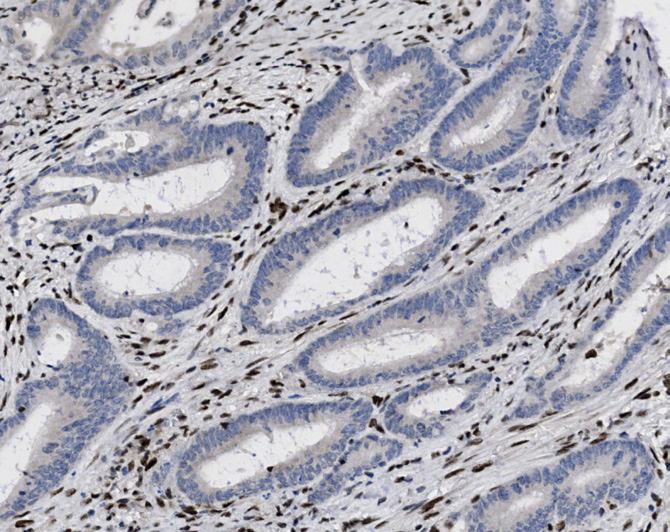
Tissue-Tek Genie® anti-MSH2
8327-M250: RTU, 1 cartridge, 250 tests; 1 unit
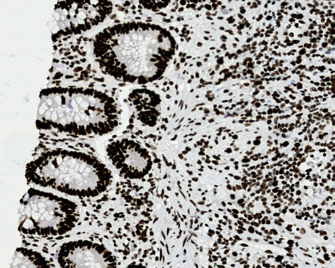
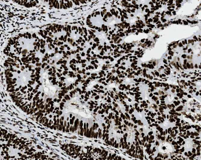
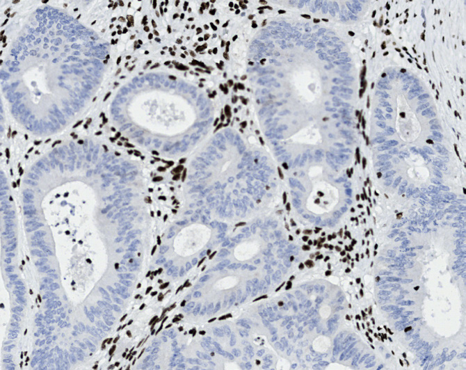
Tissue-Tek Genie® anti-MSH6
8326-M250: RTU, 1 cartridge, 250 tests; 1 unit
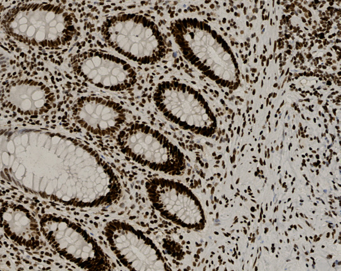
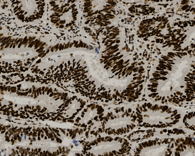
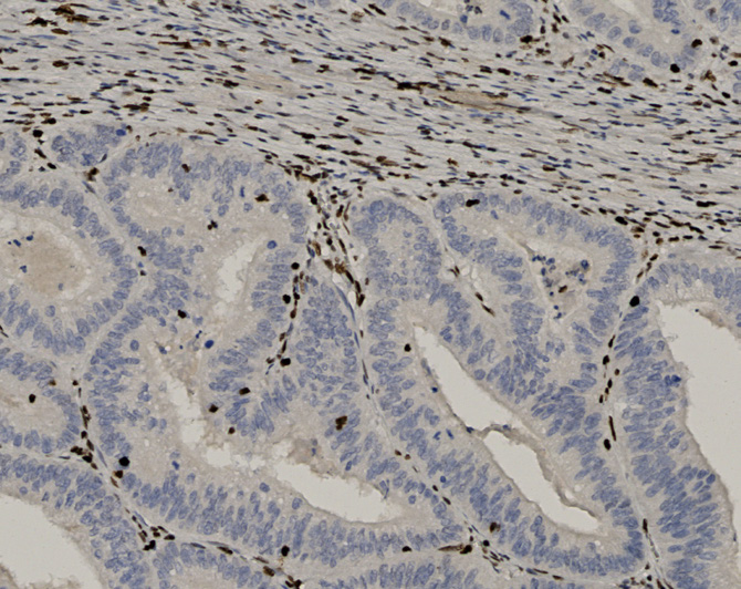
Tissue-Tek Genie® anti-MUC1
8375-M250: RTU, 250 tests, 1 cartridge; 1 unit
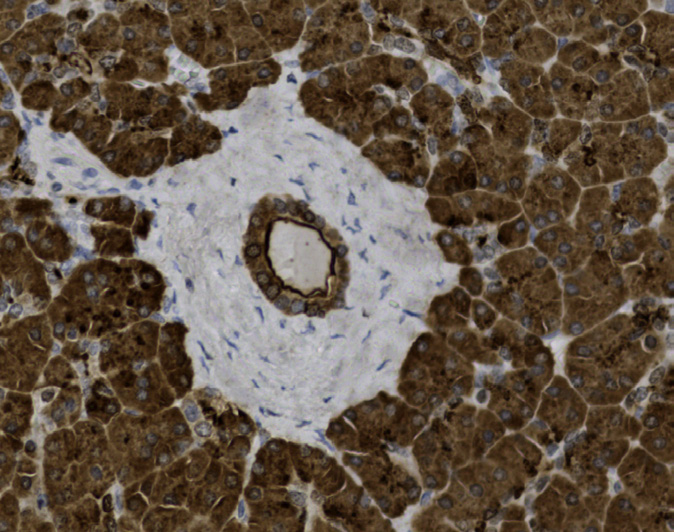
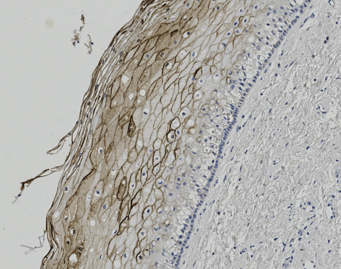
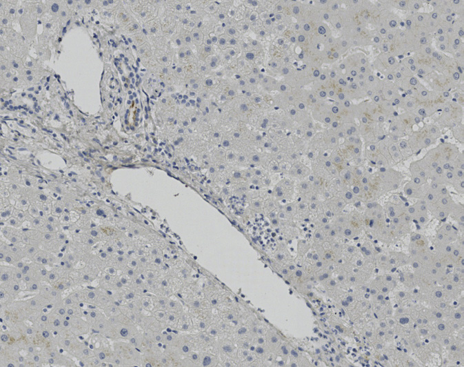
Tissue-Tek Genie® anti-MUC2
8305-M250: RTU, 250 tests, 1 cartridge; 1 unit
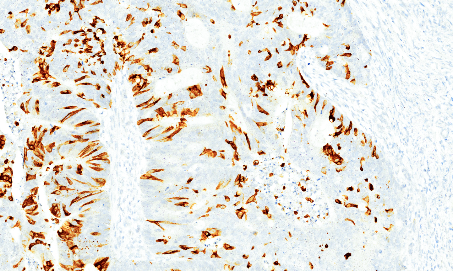
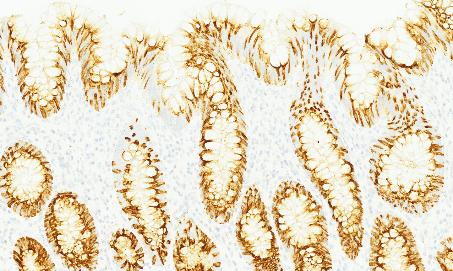
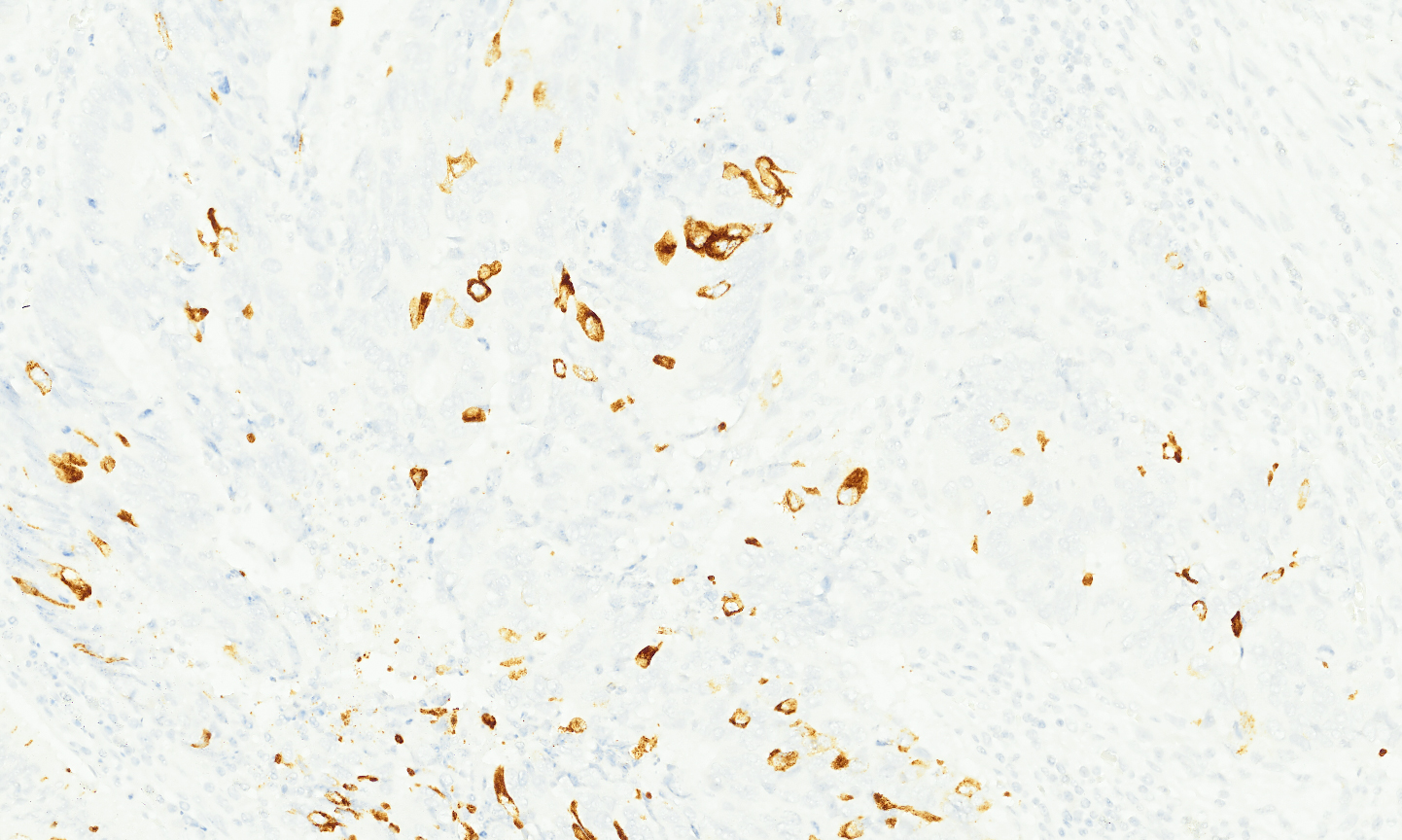
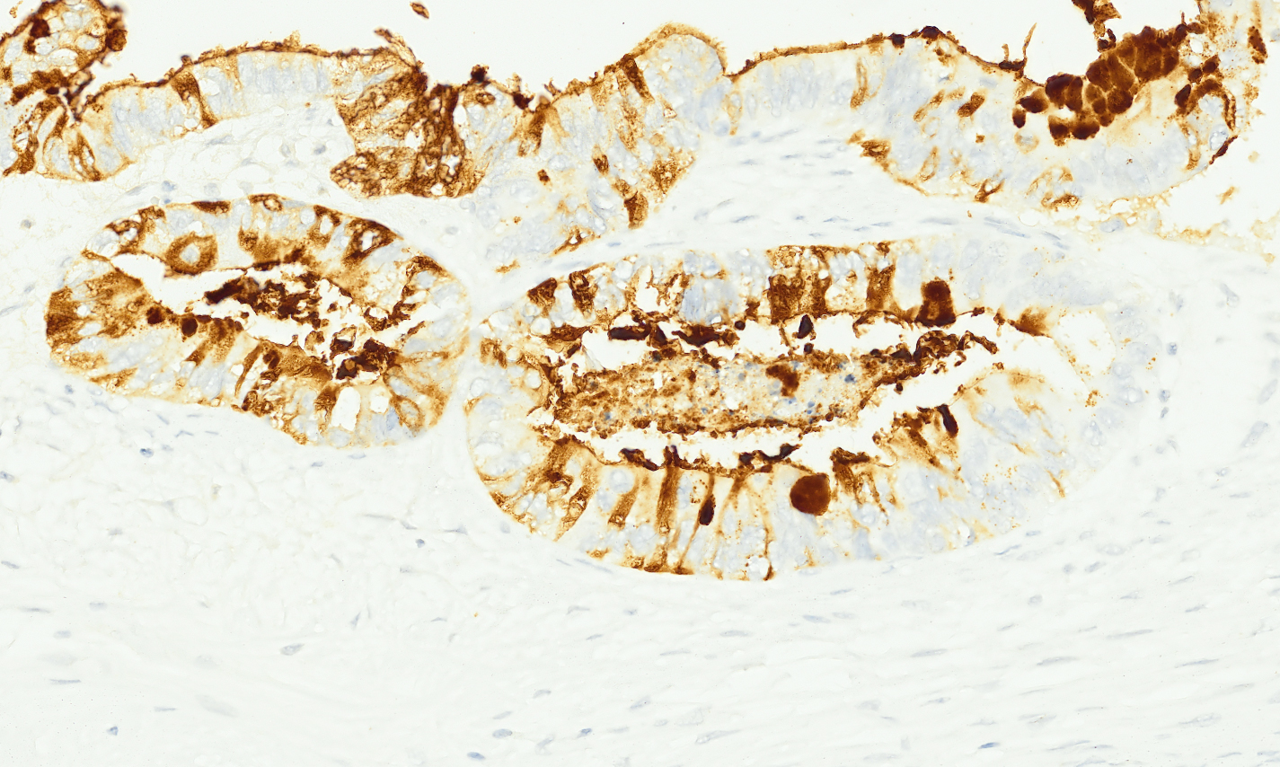
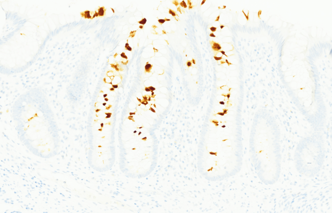
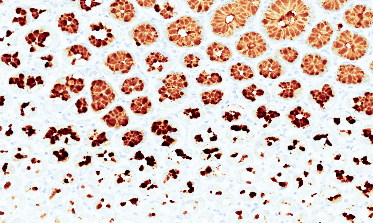
The Tissue-Tek Genie® anti-MUC2 Mouse Monoclonal Antibody [CCP58] is designed to qualitatively detect MUC2 protein in formalin-fixed, paraffin-embedded (FFPE) human tissue specimen sections by immunohistochemistry (IHC) staining on the automated Tissue-Tek Genie® Advanced Staining System. This device functions as an aid for diagnosis and shall be used by a qualified pathologist with a panel of other antibodies to identify adenocarcinoma of gastrointestinal origin and to differentiate metastatic colorectal adenocarcinoma from primary ovarian carcinoma. MUC2 is a well-established mucin marker found across a broad spectrum of gastrointestinal and other mucinous tumors. It is frequently expressed in primary GI cancers, including those of colonic, gastric, and esophageal origin. Evidence indicates that MUC2 is useful for identifying mucinous subtypes of colorectal and gastric adenocarcinomas. In contrast, non-mucinous ovarian carcinomas typically lack MUC2 expression.
Tissue-Tek Genie® anti-MUC5AC
8299-M250: RTU, 250 tests, 1 cartridge; 1 unit
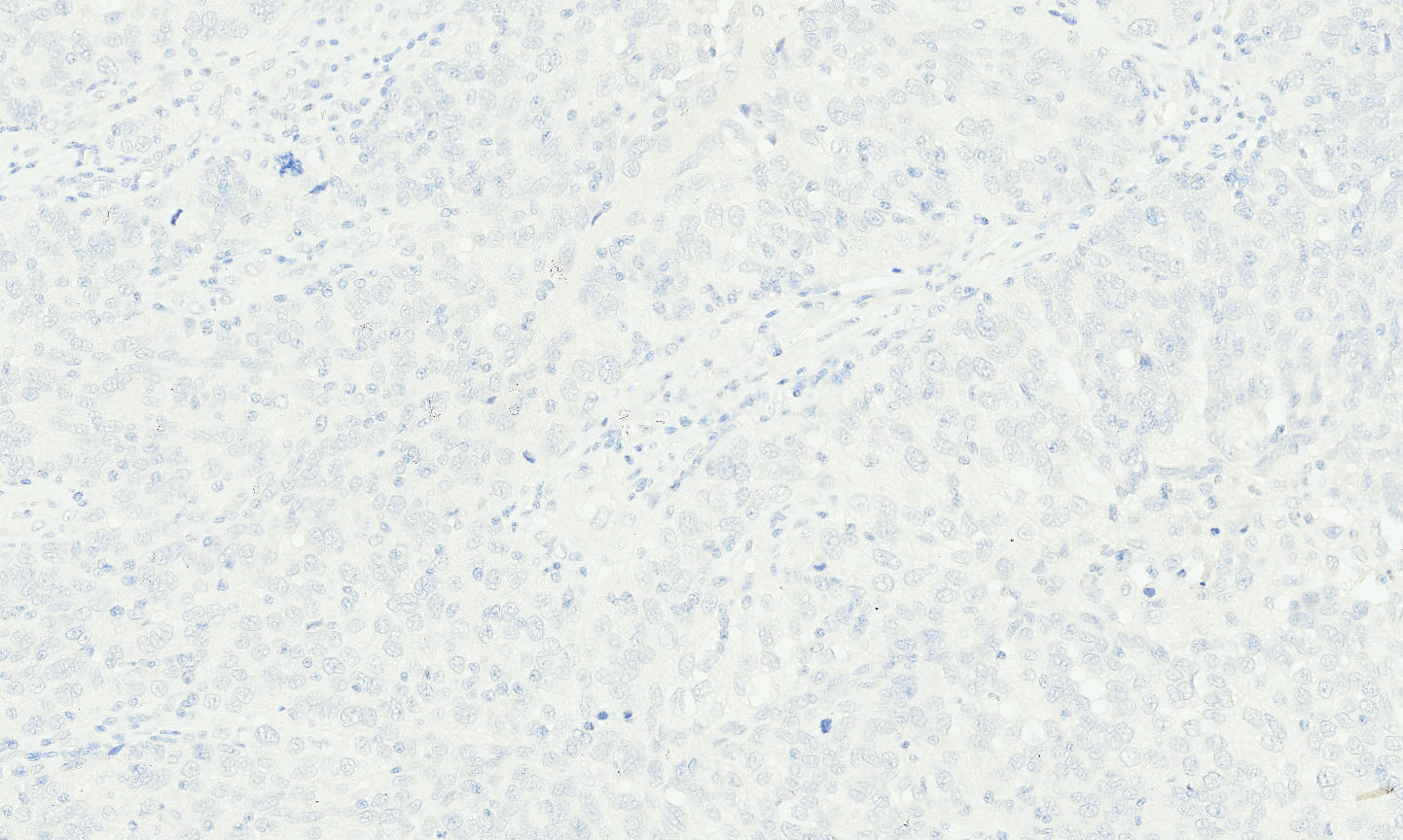
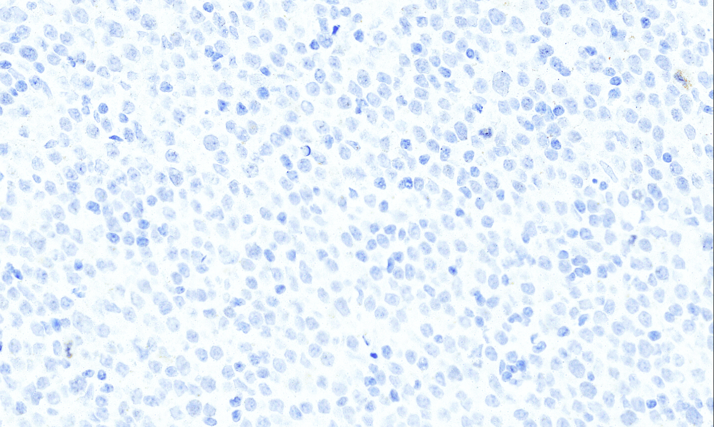
The Tissue-Tek Genie® anti-MUC5AC Mouse Monoclonal Antibody [Nd-2] is designed to qualitatively detect MUC5AC protein in formalin-fixed, paraffin-embedded (FFPE) human tissue specimen sections by immunohistochemistry (IHC) staining on the automated Tissue-Tek Genie® Advanced Staining System. This device functions as an aid for diagnosis and shall be used by a qualified pathologist with a panel of other antibodies to differentiate pancreatic intraepithelial neoplasia and invasive pancreatic adenocarcinoma from benign/reactive pancreatic conditions. MUC5AC is a widely recognized mucin biomarker. Its expression is frequently upregulated in pancreatic intraepithelial neoplasia and is commonly observed in pancreatic ductal adenocarcinoma. In contrast, MUC5AC expression is typically absent in sarcomas and lymphomas.
Tissue-Tek Genie® anti-MUM-1
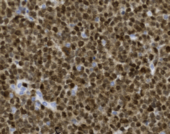
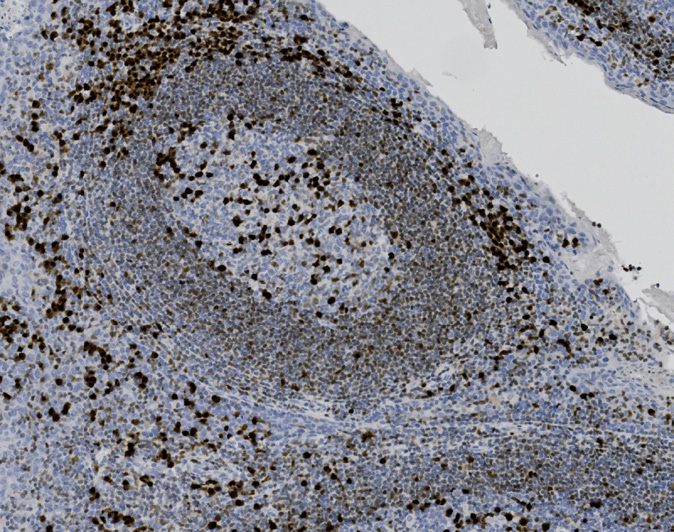
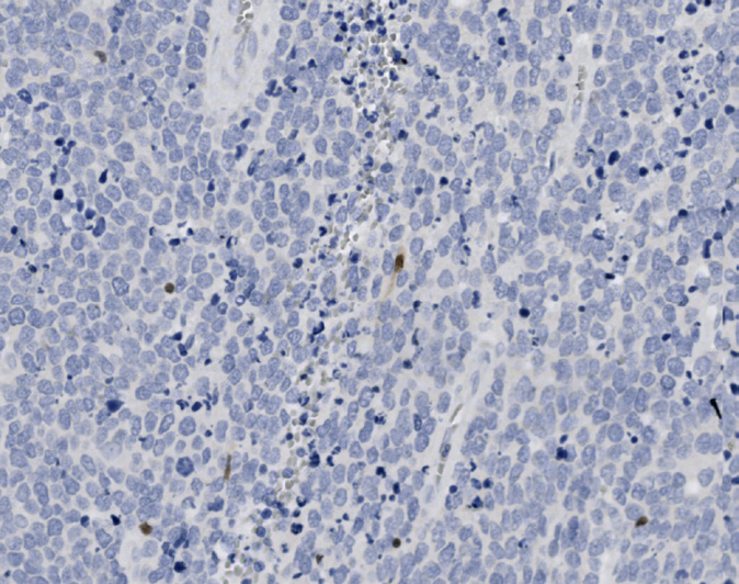
8329-M250: RTU, 250 tests, 1 cartridge; 1 unit
Tissue-Tek Genie® anti-Myeloperoxidase (MPO)
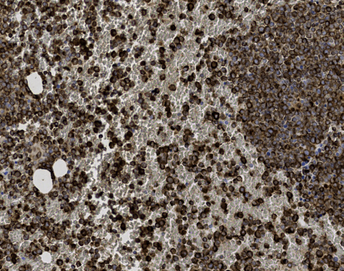
8407-M250: RTU, 250 tests, 1 cartridge; 1 unit
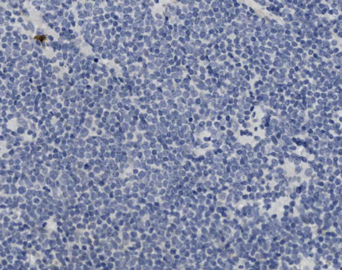
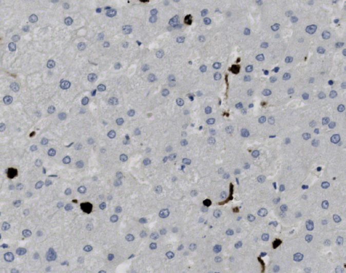
Tissue-Tek Genie® anti-Myosin Smooth Muscle
8517-M250: RTU, 250 tests, 1 cartridge; 1 unit
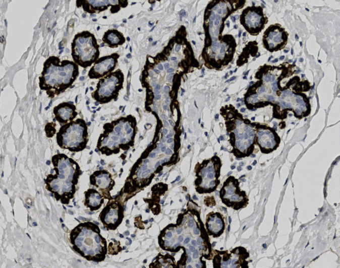
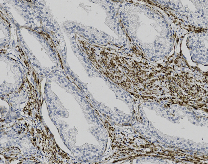
Tissue-Tek Genie® anti-Napsin A
8331-M250: RTU, 250 tests, 1 cartridge; 1 unit
Tissue-Tek Genie® anti-Neurofilament
8289-M250: RTU, 250 tests, 1 cartridge; 1 unit
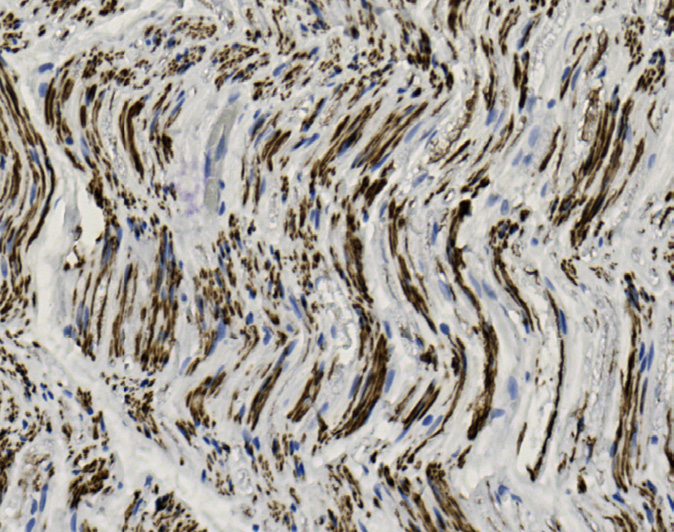
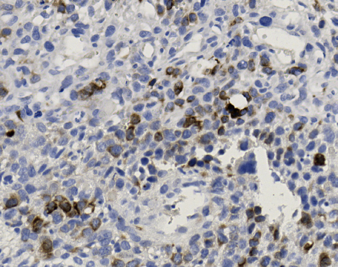
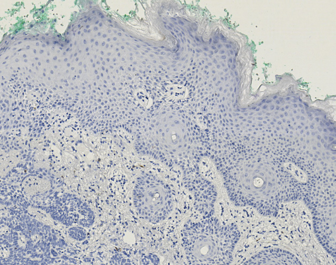
Tissue-Tek Genie® anti-Neuron Specific Enolase
8447-M250: RTU, 250 tests, 1 cartridge; 1 unit
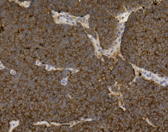
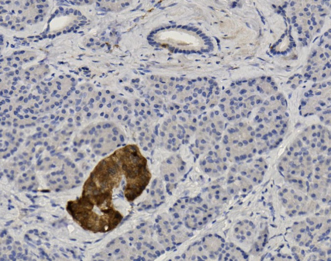
Tissue-Tek Genie® anti-NKX3.1
8320-M250: RTU, 250 tests, 1 cartridge; 1 unit
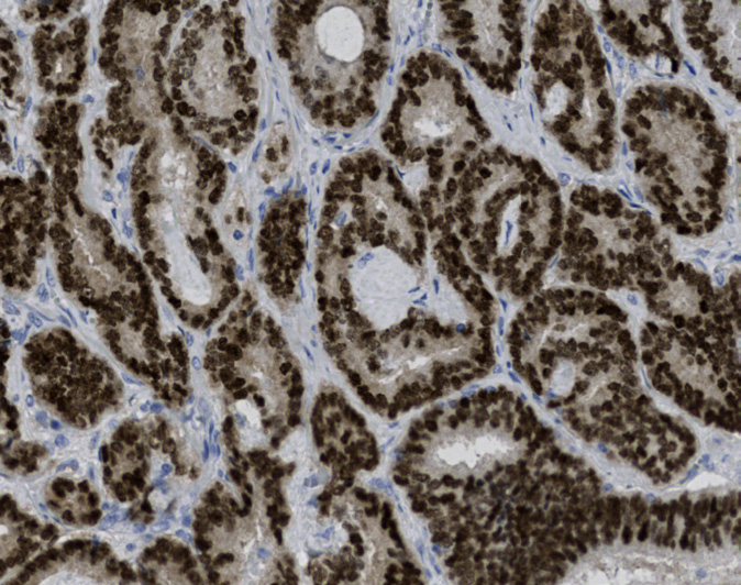
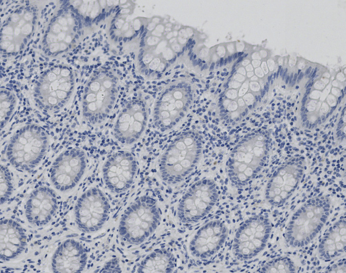
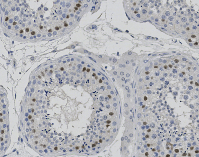
Tissue-Tek Genie® anti-OCT.2
8334-M250: RTU, 250 tests, 1 cartridge; 1 unit
Tissue-Tek Genie® anti-OCT.3/4
8288-M250: RTU, 250 tests, 1 cartridge; 1 unit
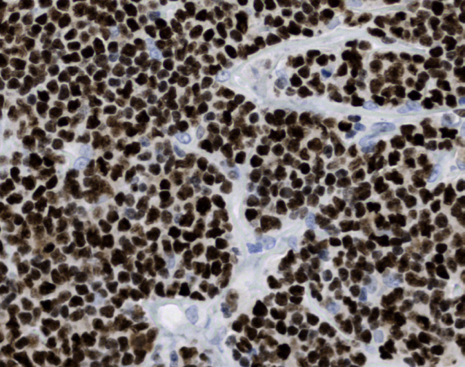
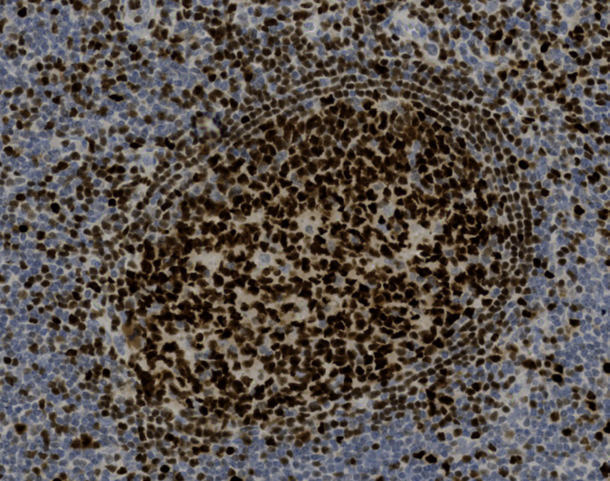
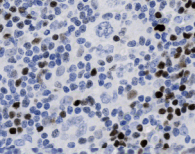
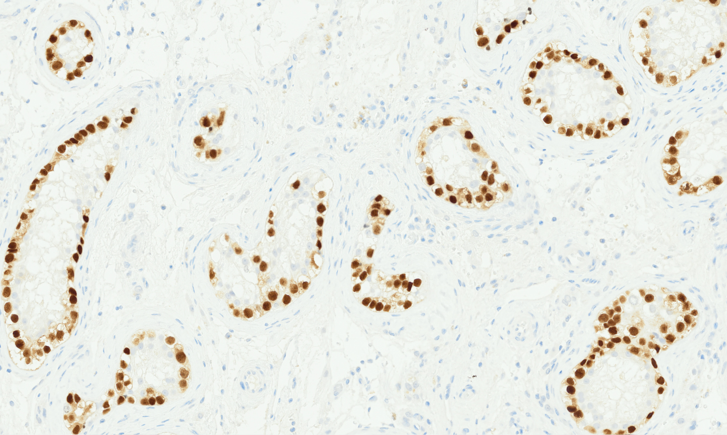
The Tissue-Tek Genie® anti-OCT3/4 Mouse Monoclonal Antibody [C-10] is designed to qualitatively detect OCT3/4 protein in formalin-fixed, paraffin-embedded (FFPE) human tissue specimen sections by immunohistochemistry (IHC) staining on the automated Tissue-Tek Genie® Advanced Staining System. OCT3/4 IHC is used as a diagnostic tool with a panel of other antibodies in identifying germ cell tumors and distinguishing them from other neoplasms. In tumor tissues, OCT3/4 expression varies by germ cell tumor (GCT) type and has been detected in the neoplastic cells of seminoma, dysgerminoma, embryonal carcinoma, and germ cell neoplasia in situ (GCNIS). However, OCT3/4 is absent in yolk sac tumor, teratoma and choriocarcinoma.
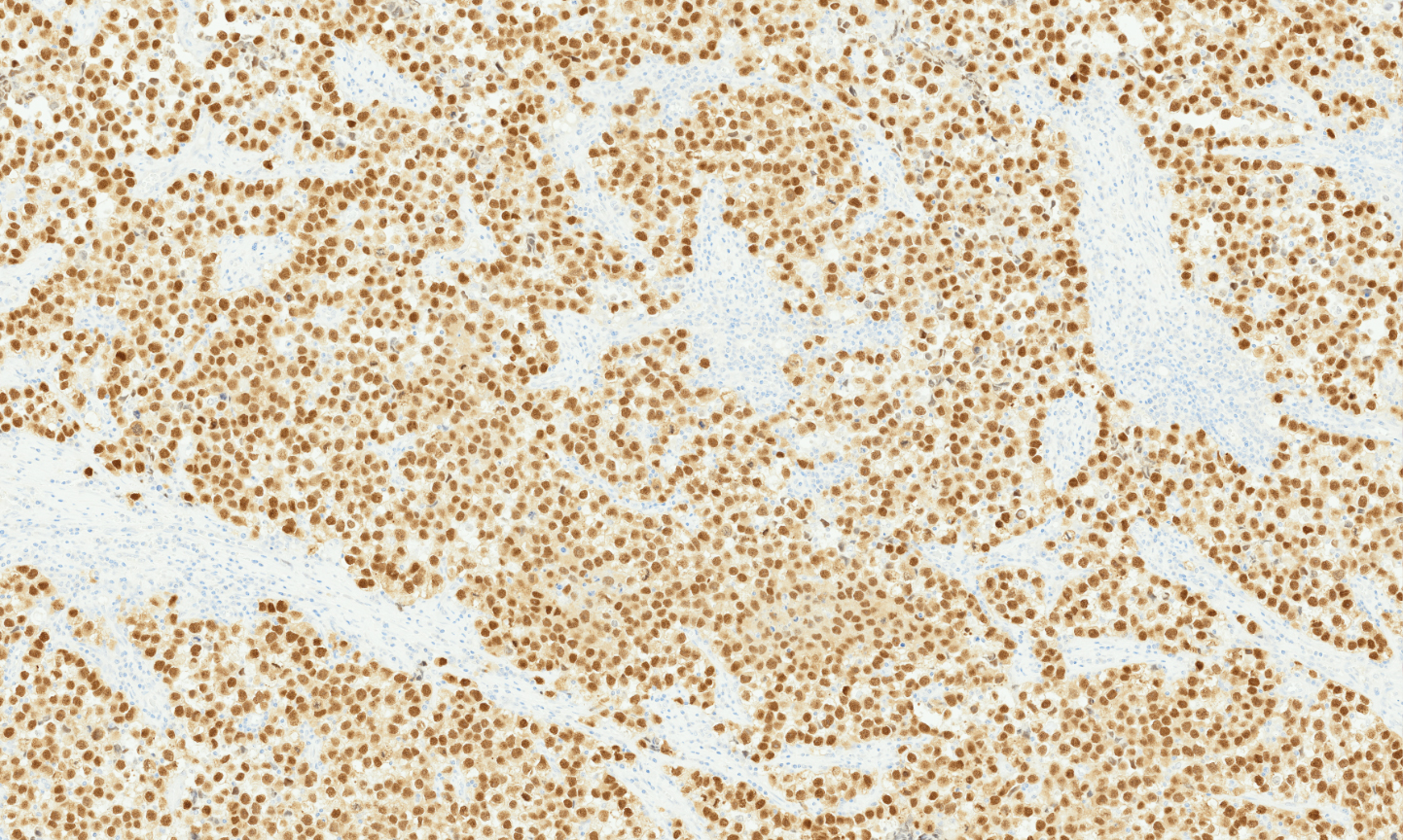
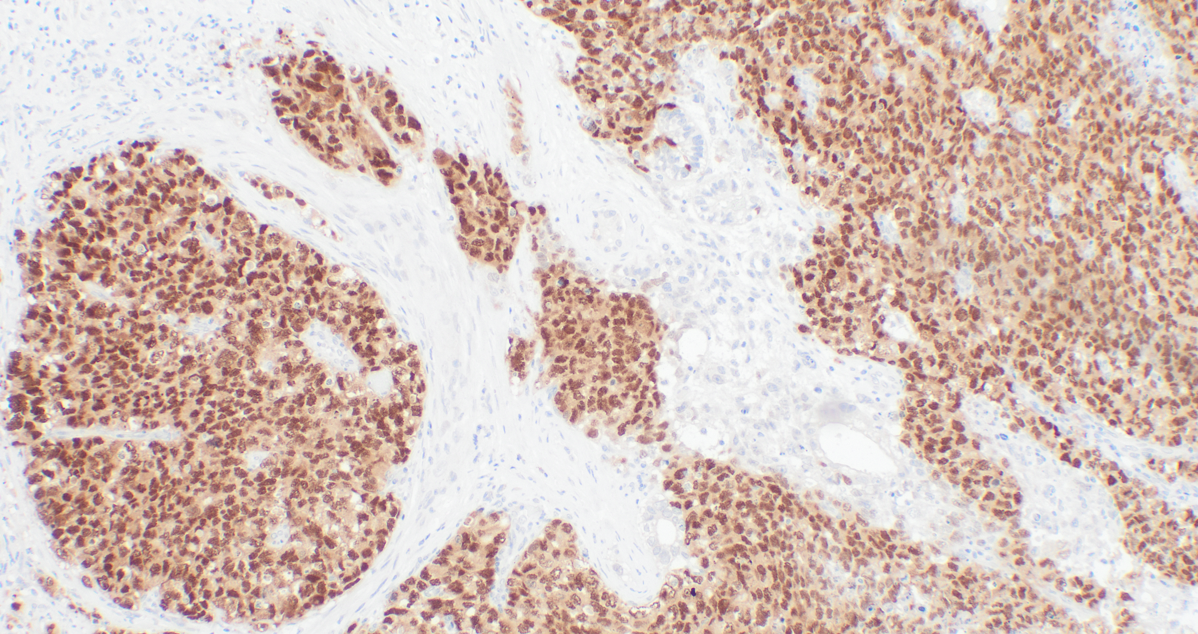
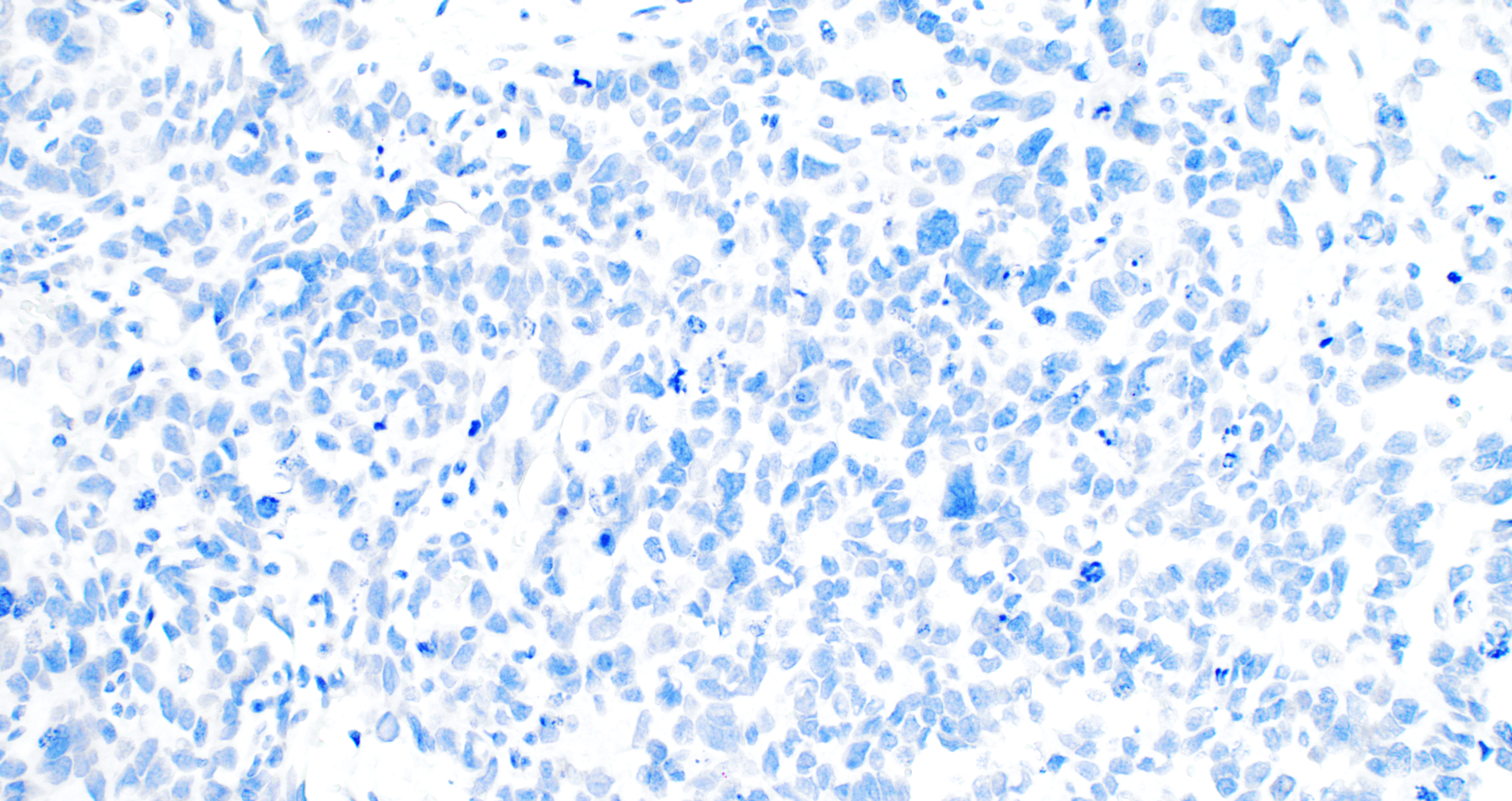
Tissue-Tek Genie® anti-p40
8341-M250: RTU, 250 tests, 1 cartridge; 1 unit
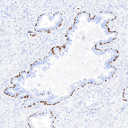
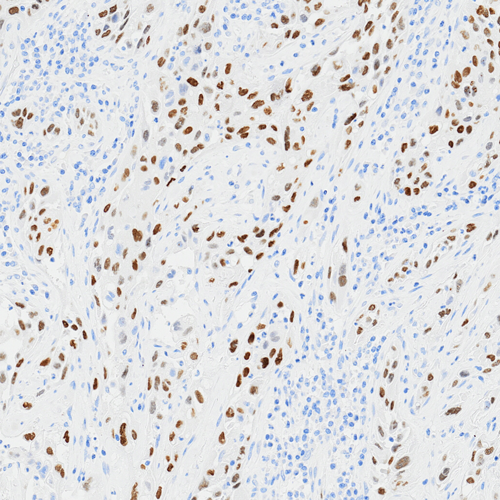
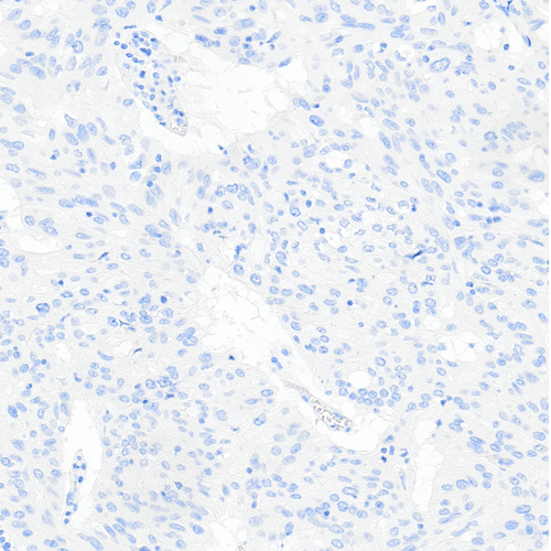
The Tissue-Tek Genie® anti-p40 Mouse Monoclonal Antibody [BC28] is designed to qualitatively detect p40 protein in formalin-fixed, paraffin-embedded (FFPE) human tissue specimen sections by immunohistochemistry (IHC) staining on the automated Tissue-Tek Genie® Advanced Staining System.
This device functions as an aid for diagnosis and shall be used by a qualified pathologist with a panel of other antibodies to differentiate lung squamous cell carcinoma from lung adenocarcinoma and to identify urothelial neoplasms.
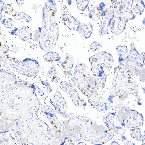
Tissue-Tek Genie® anti-p40 / Myosin Smooth Muscle Antibody Cocktail
8342-M250: RTU, 250 tests, 1 cartridge; 1 unit
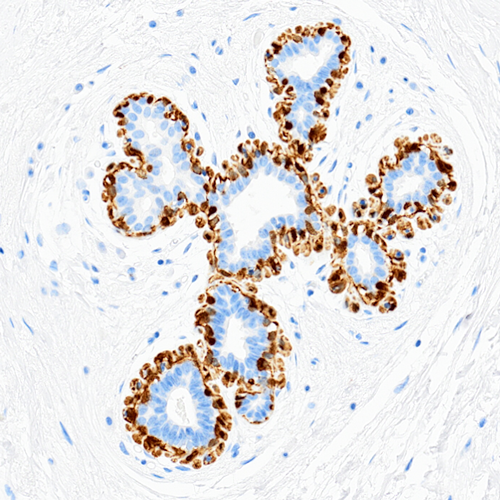
The Tissue-Tek Genie® anti-p40/Myosin Smooth Muscle Antibody Cocktail [BC28/SMMS-1] is designed to qualitatively detect p40 and myosin smooth muscle proteins in formalin-fixed, paraffin-embedded (FFPE) human tissue specimen sections by immunohistochemistry (IHC) staining on the automated Tissue-Tek Genie® Advanced Staining System.
This device functions as an aid for diagnosis and shall be used by a qualified pathologist with a panel of other antibodies to differentiate invasive breast carcinoma from noninvasive breast proliferations such as ductal carcinoma in situ (DCIS).
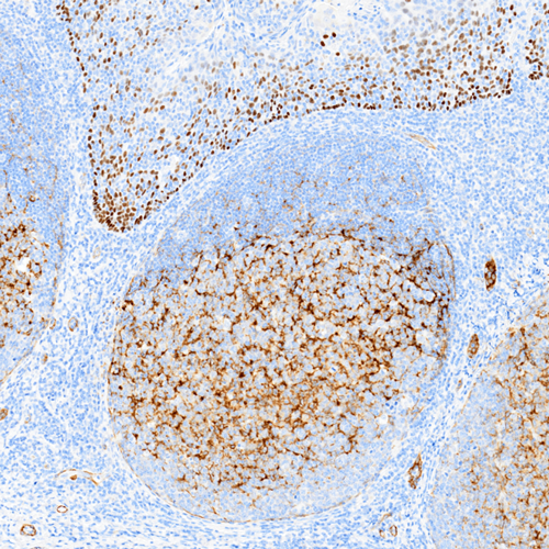
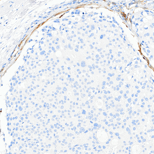
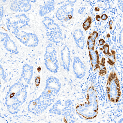
Tissue-Tek Genie® anti-p53
8477-M250: RTU, 1 cartridge, 250 tests; 1 unit
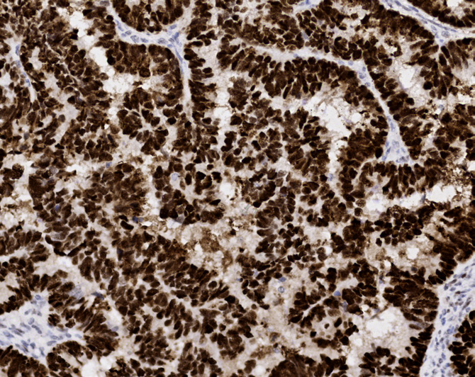
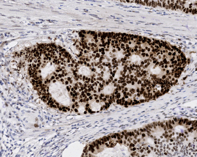
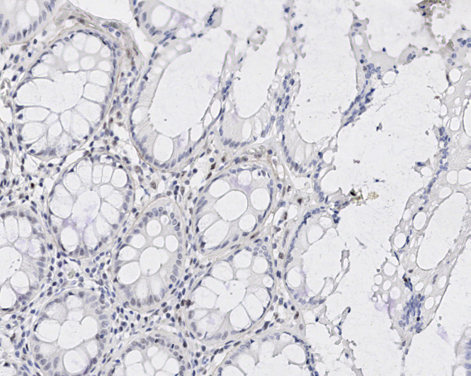
Tissue-Tek Genie® anti-p63
8312-M250: RTU, 250 tests, 1 cartridge; 1 unit
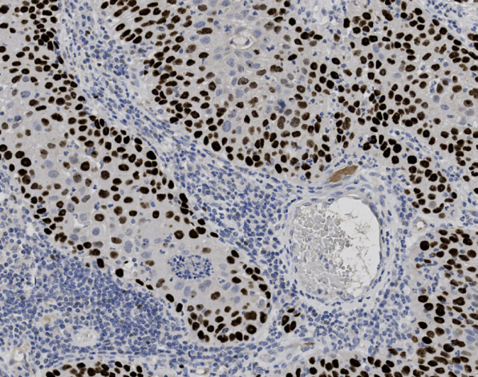
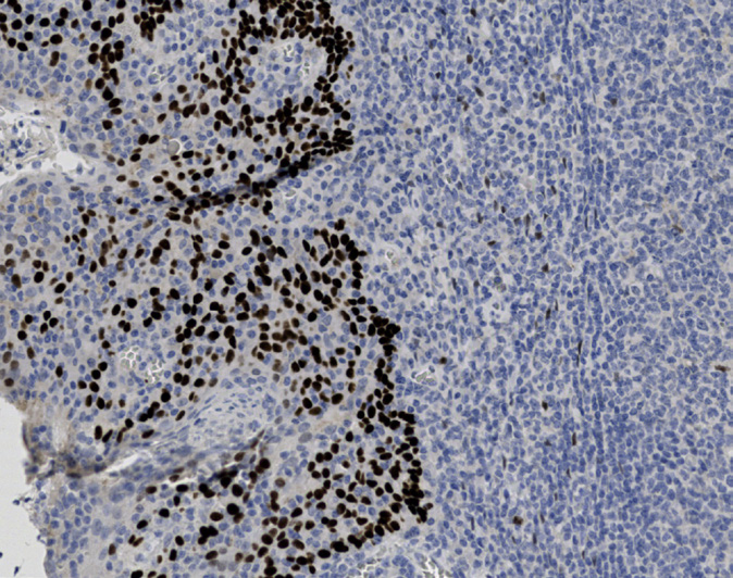
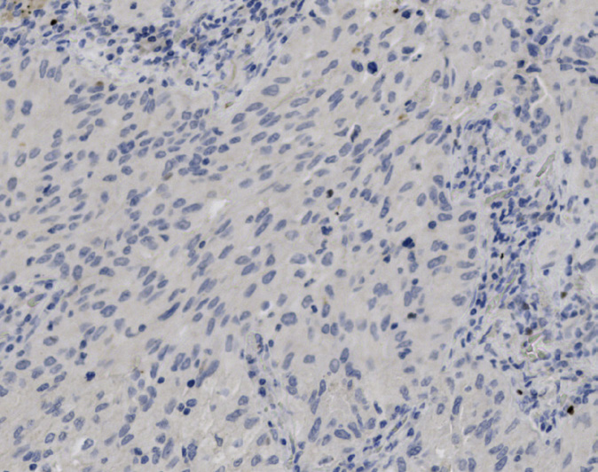
Tissue-Tek Genie® anti-p120
8373-M250: RTU, 250 tests, 1 cartridge; 1 unit
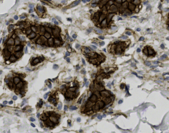
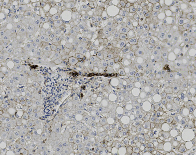
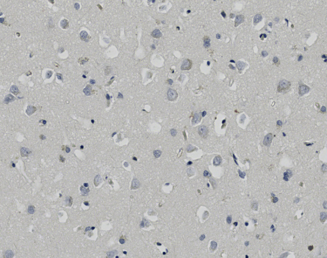
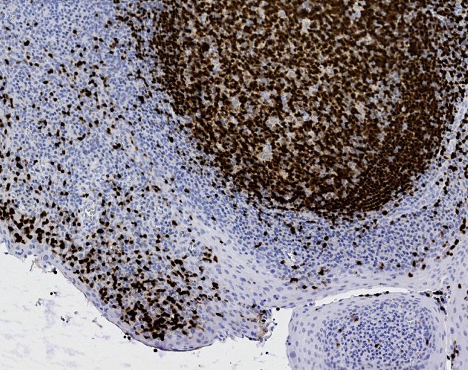
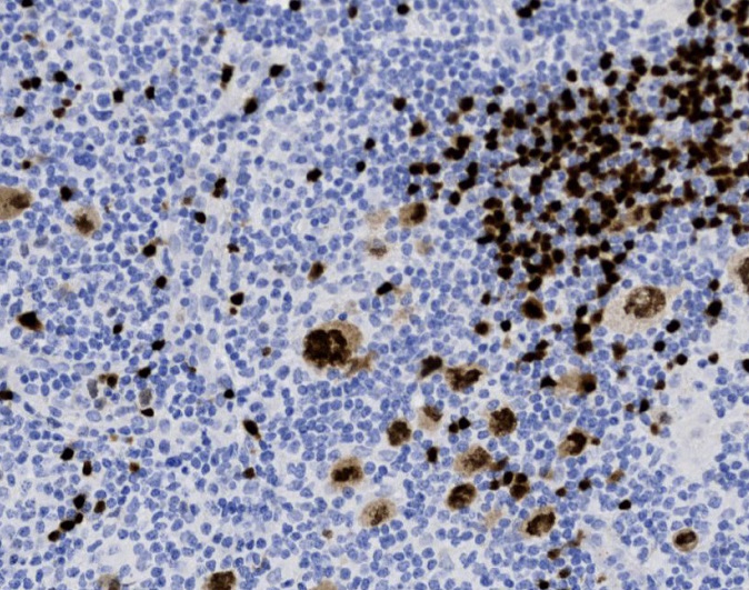
Tissue-Tek Genie® anti-PAX5
8500-M250: RTU, 250 tests, 1 cartridge; 1 unit
Tissue-Tek Genie® anti-PAX8
8257-M250: RTU, 250 tests, 1 cartridge; 1 unit
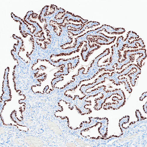
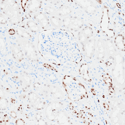
The Tissue-Tek Genie® anti-PAX8 Rabbit Monoclonal Antibody [RM436] is designed to qualitatively detect PAX8 protein in formalin-fixed, paraffin-embedded (FFPE) human tissue specimen sections by immunohistochemistry (IHC) staining on the automated Tissue-Tek Genie® Advanced Staining System.
This device functions as an aid for diagnosis and shall be used by a qualified pathologist with a panel of other antibodies to identify renal cell carcinoma and ovarian non-mucinous carcinoma.
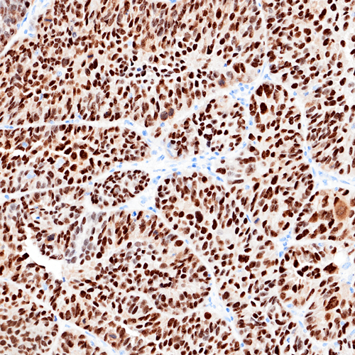
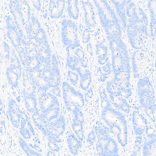
Tissue-Tek Genie® anti-PD-1
8287-M250: RTU, 250 tests, 1 cartridge; 1 unit
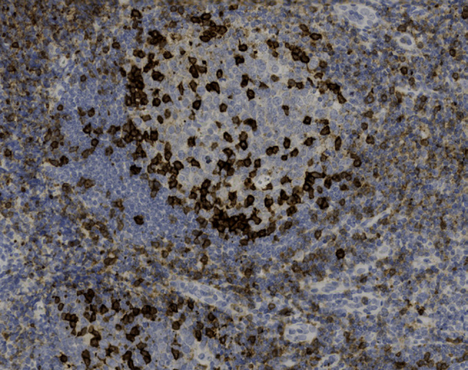
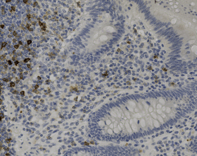
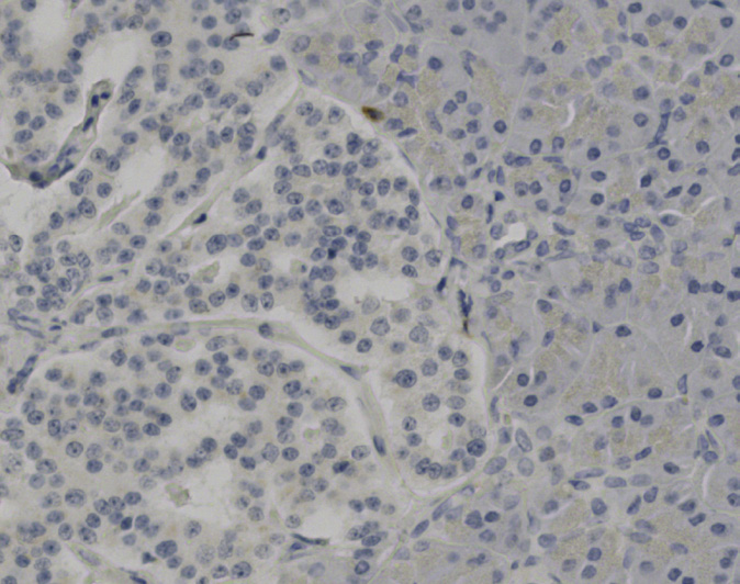
Tissue-Tek Genie® anti-Perforin
8236-M250: RTU, 250 tests, 1 cartridge; 1 unit
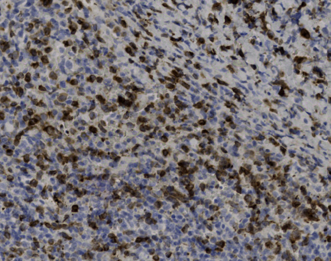
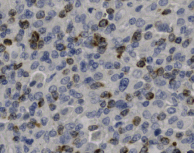
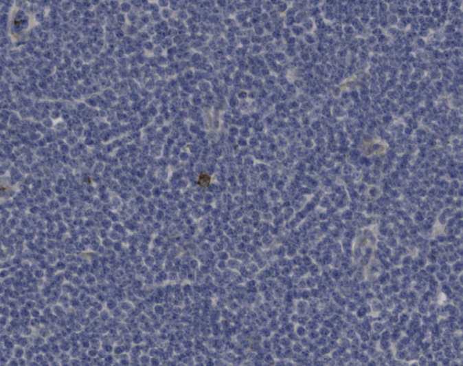
Tissue-Tek Genie® anti-PLAP
8235-M250: RTU, 250 tests, 1 cartridge; 1 unit
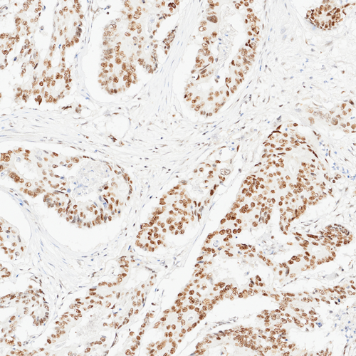
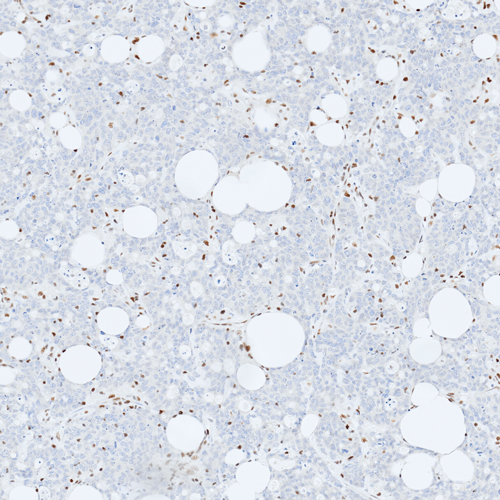
Tissue-Tek Genie® anti-PMS2
8328-M250: RTU, 250 tests, 1 cartridge; 1 unit
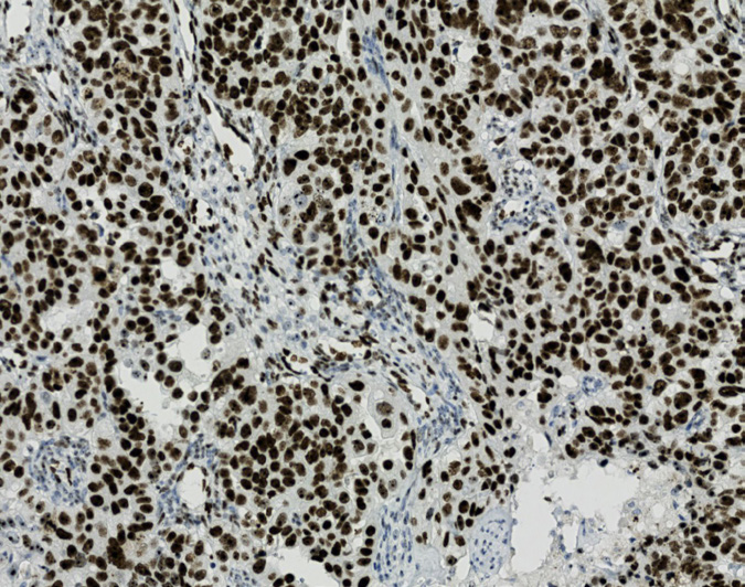
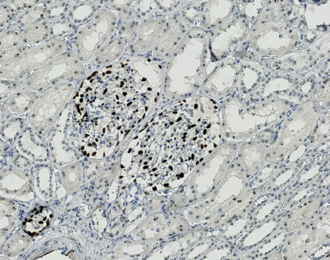
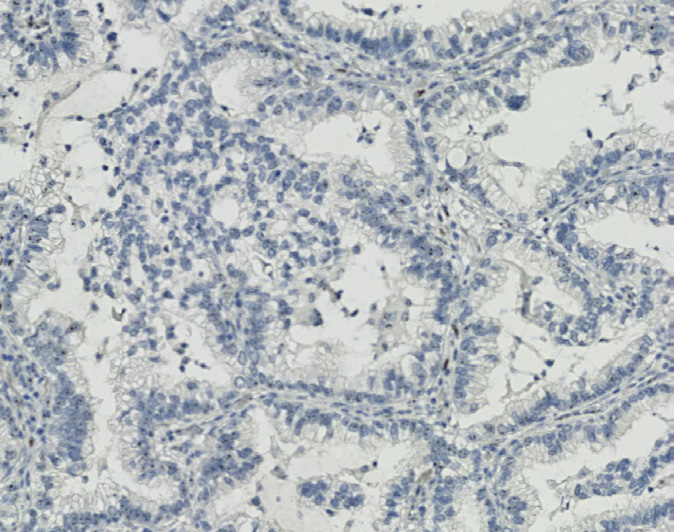
The Tissue-Tek Genie® anti-PMS2 Rabbit Monoclonal Antibody [EP51] is designed to qualitatively detect PMS2 protein in formalin-fixed, paraffin-embedded (FFPE) human tissue specimen sections by immunohistochemistry (IHC) staining on the automated Tissue-Tek Genie® Advanced Staining System.
This device functions as an aid for diagnosis and shall be used by a qualified pathologist with a panel of other antibodies to classify colorectal cancers.
Tissue-Tek Genie® anti-Podoplanin
8515-M250: RTU, 250 tests, 1 cartridge; 1 unit
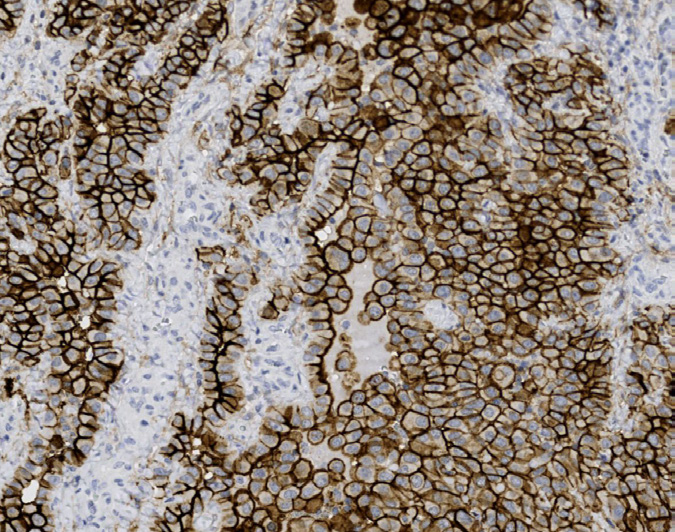
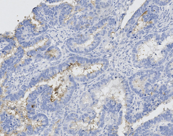
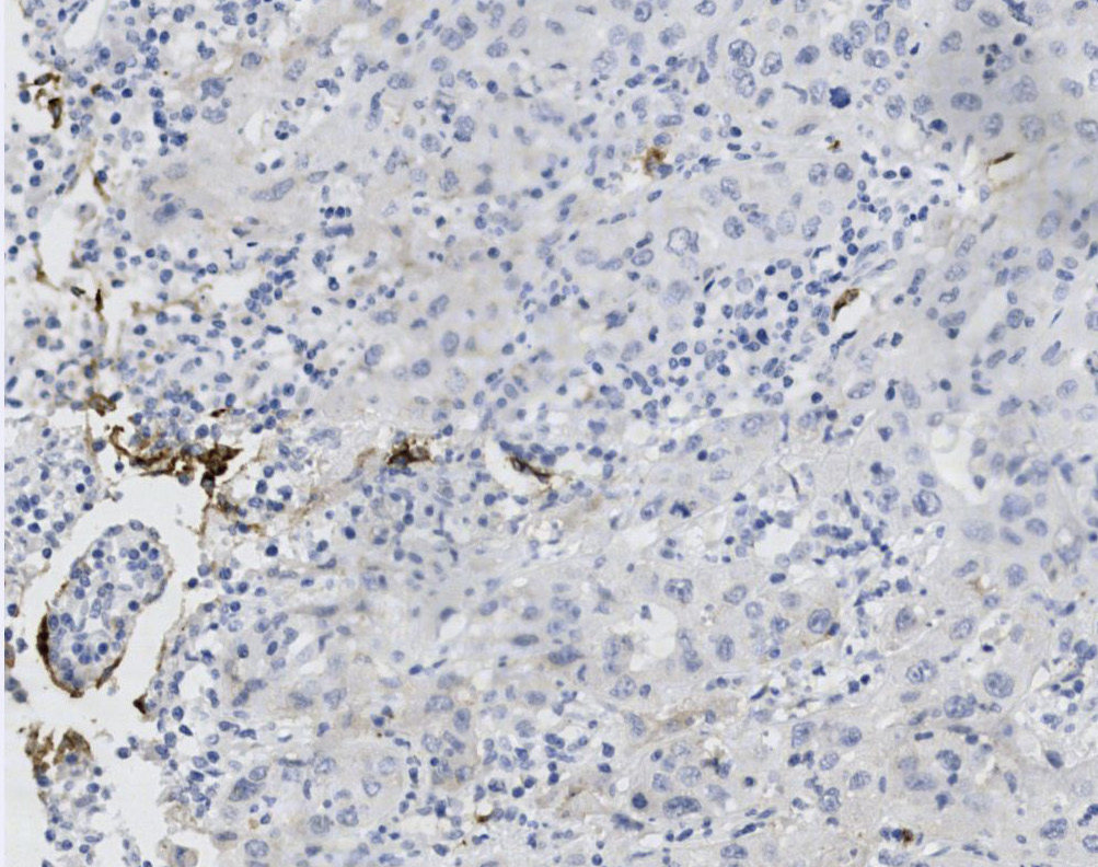
Tissue-Tek Genie® anti-Prostate Specific Antigen
8244-M250: RTU, 250 tests, 1 cartridge; 1 unit
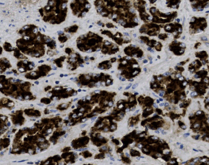
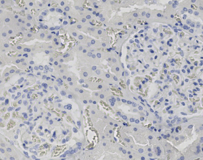
Tissue-Tek Genie® anti-Prostein
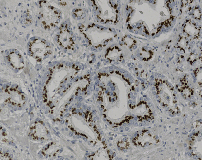
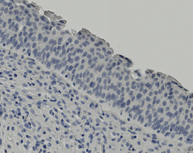
8519-M250: RTU, 250 tests, 1 cartridge; 1 unit
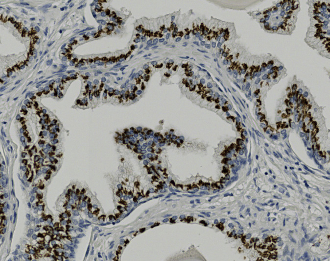
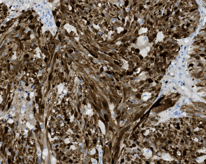
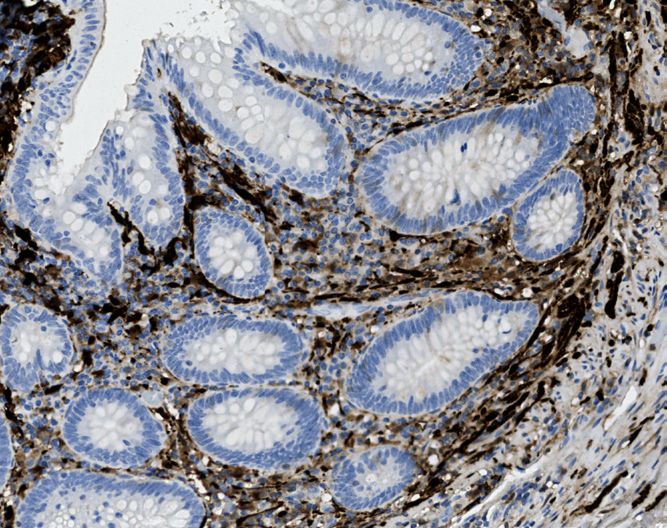
Tissue-Tek Genie® anti-S100B
8442-M250: RTU, 1 cartridge, 250 tests; 1 unit
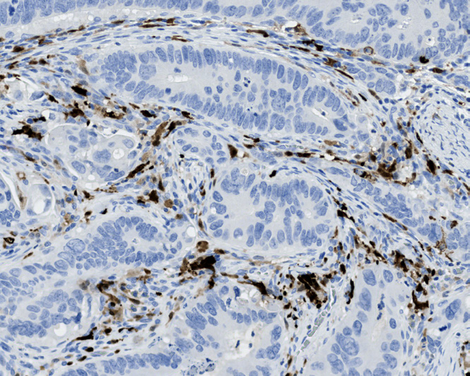
Tissue-Tek Genie® anti-SALL4
8358-M250: RTU, 250 tests, 1 cartridge; 1 unit
The Tissue-Tek Genie® anti-SALL4 Mouse Monoclonal Antibody [6E3] is designed to qualitatively detect SALL4 protein in formalin-fixed, paraffin-embedded (FFPE) human tissue specimen sections by immunohistochemistry (IHC) staining on the automated Tissue-Tek Genie® Advanced Staining System. This device functions as an aid for diagnosis and shall be used by a qualified pathologist with a panel of other antibodies to identify germ cell tumors such as seminoma, dysgerminoma, embryonal carcinoma, yolk sac tumor, and germ cell neoplasia in situ (GCNIS) and distinguish them from other neoplasms. SALL4 expression is variably detected across germ cell tumors (GCTs) but is consistently observed in seminomas, dysgerminomas, embryonal carcinomas, yolk sac tumors, and germ cell neoplasia in situ (GCNIS). In contrast, its expression is more heterogeneous in choriocarcinomas and immature teratomas.
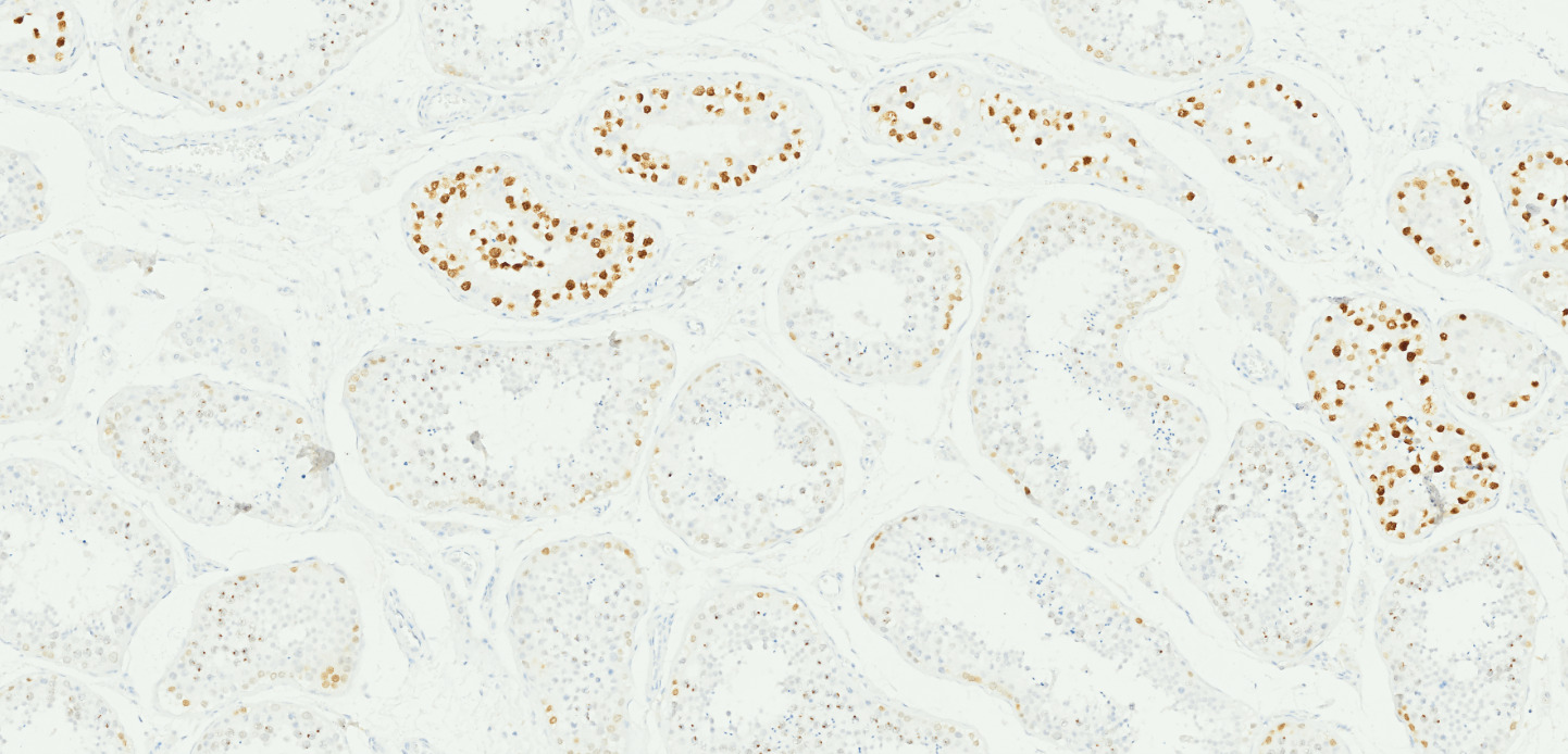
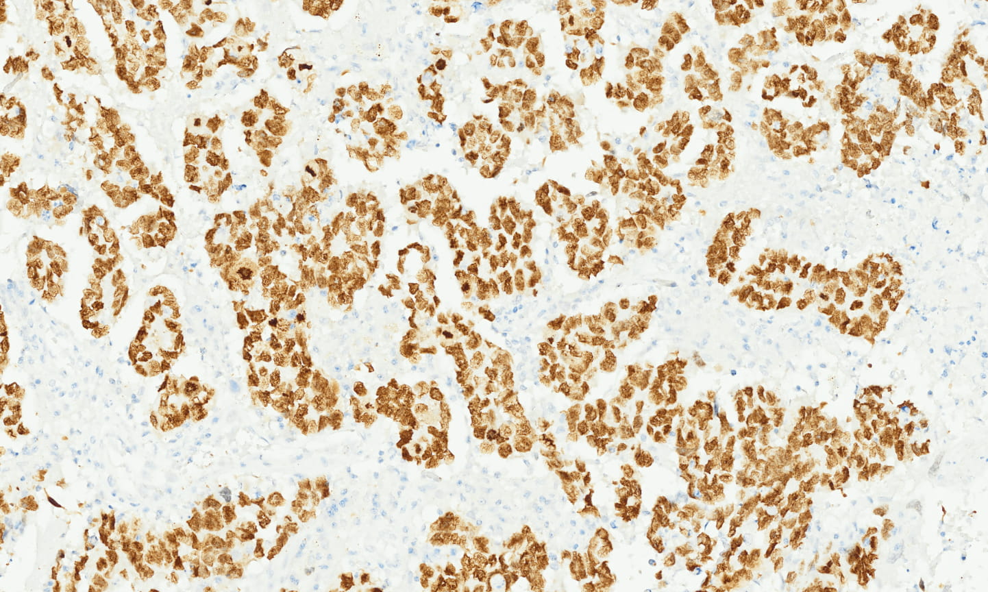
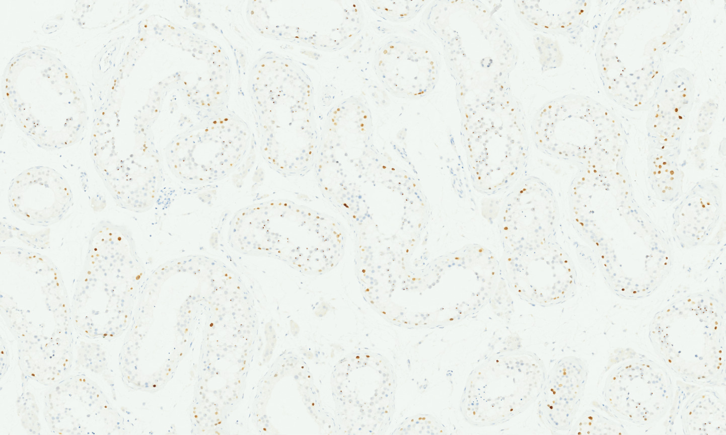
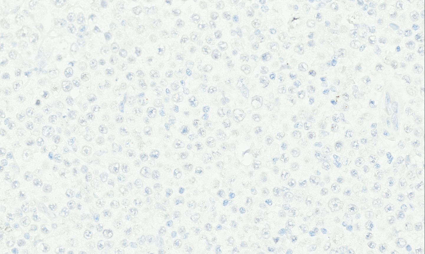
Tissue-Tek Genie® anti-SATB2
8356-M250: RTU, 250 tests, 1 cartridge; 1 unit
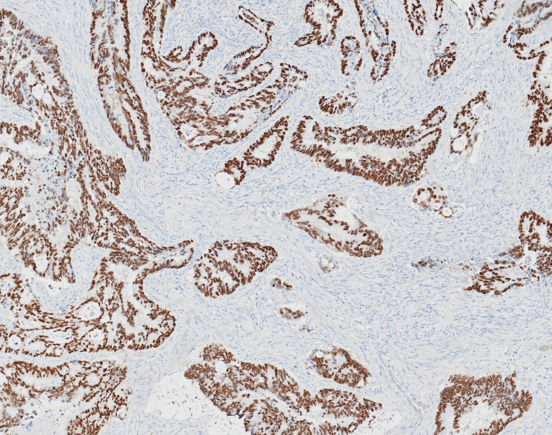
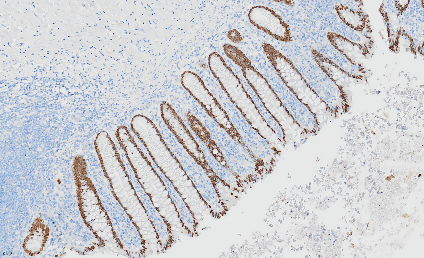
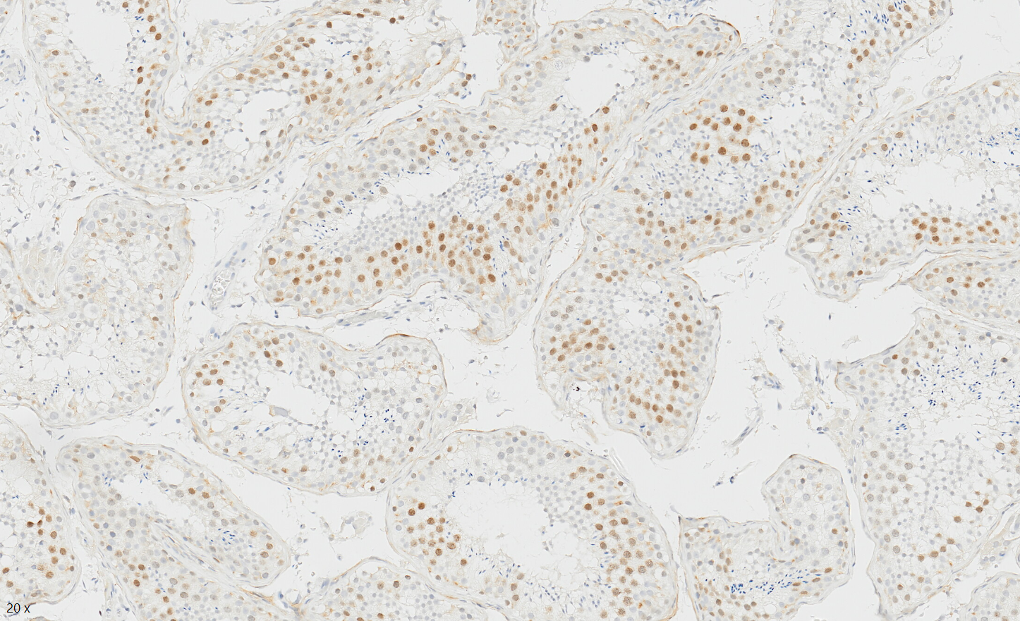
Special AT-rich sequence-binding protein 2 (SATB2) is a nuclear transcription factor that is strongly expressed in the epithelium of the lower gastrointestinal tract (GI) including the colon, appendix, and rectum.
Tissue-Tek Genie® anti-SOX2
8228-M250: RTU, 250 tests, 1 cartridge; 1 unit
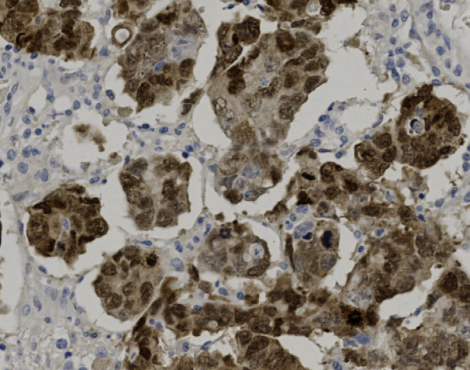
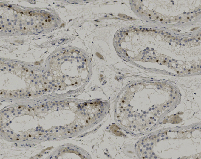
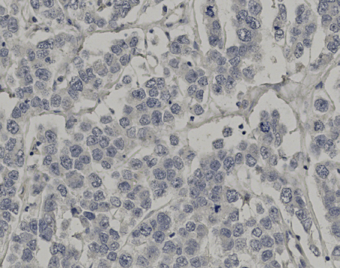
Tissue-Tek Genie® anti-SOX10
8264-M250: RTU, 1 cartridge, 250 tests; 1 unit
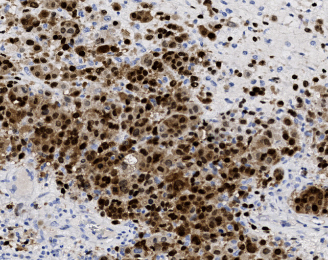
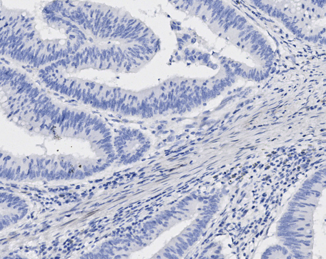
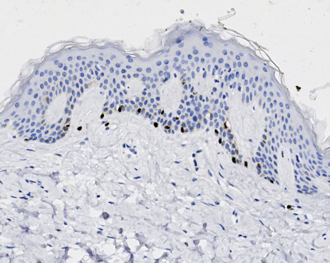
Tissue-Tek Genie® anti-SOX11
8521-M250: RTU, 250 tests, 1 cartridge; 1 unit
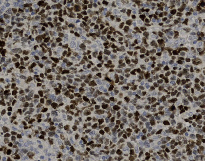
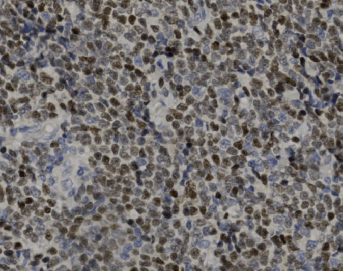
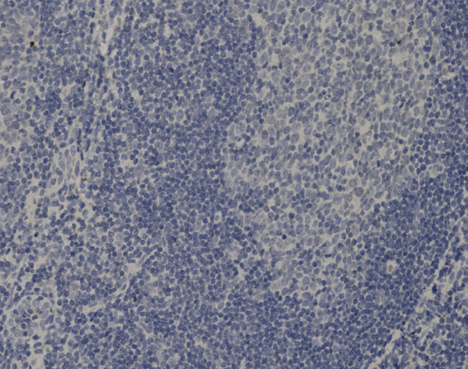
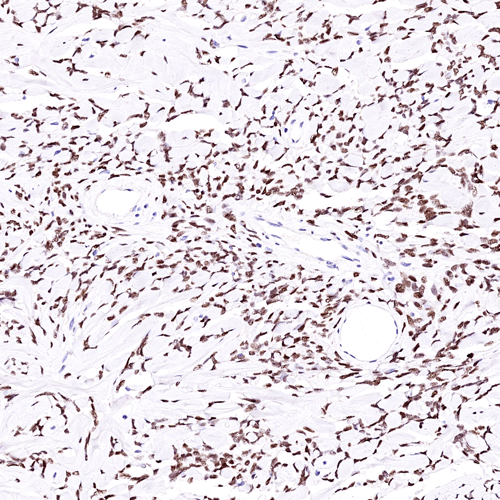
Tissue-Tek Genie® anti-STAT6
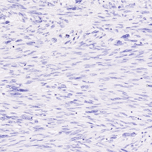
8387-M250: RTU, 250 tests, 1 cartridge; 1 unit
The Tissue-Tek Genie® anti-STAT6 Rabbit Monoclonal Antibody [EP325] is designed to qualitatively detect STAT6 protein in formalin-fixed, paraffin-embedded (FFPE) human tissue specimen sections by immunohistochemistry (IHC) staining on the automated Tissue-Tek Genie® Advanced Staining System.
This device functions as an aid for diagnosis and shall be used by a qualified pathologist with a panel of other antibodies to identify solitary fibrous tumors.
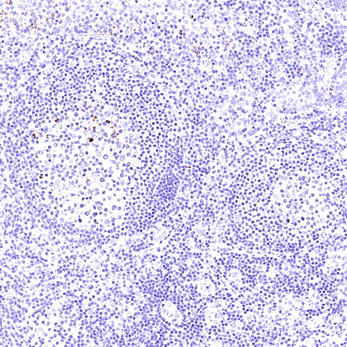
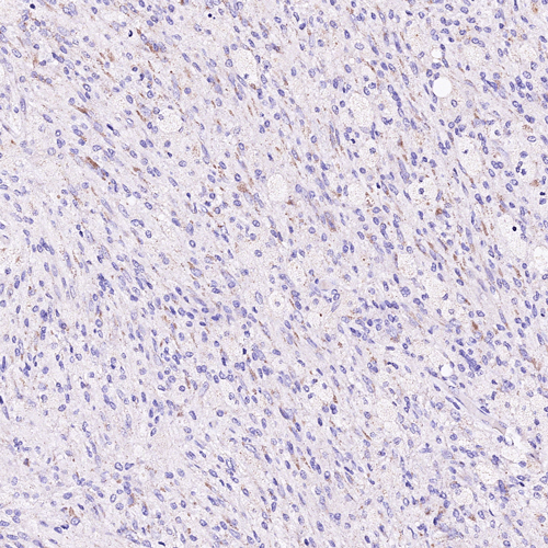
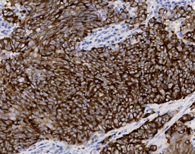
Tissue-Tek Genie® anti-Synaptophysin
8453-M250: RTU, 1 cartridge, 250 tests; 1 unit
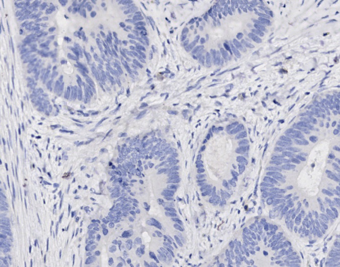
Tissue-Tek Genie® anti-Terminal Deoxyribonucleotidyl Transferase
8243-M250: RTU, 250 tests, 1 cartridge; 1 unit
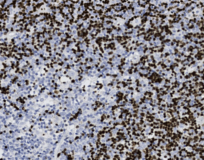
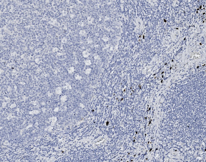
Tissue-Tek Genie® anti-Thyroid Transcription Factor-1
8224-M250: RTU, 1 cartridge, 250 tests; 1 unit
and certain areas of brain.
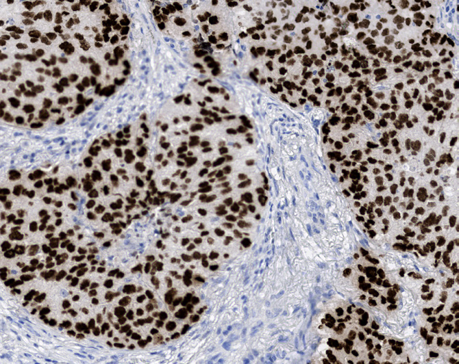
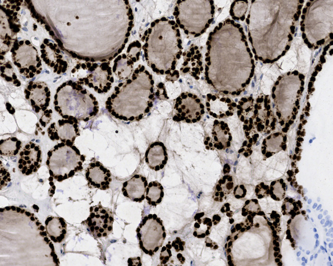
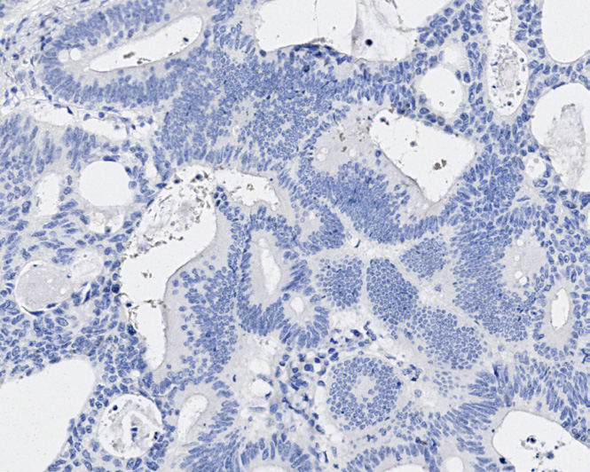
Tissue-Tek Genie® anti-TRPS1
8359-M250: RTU, 250 tests, 1 cartridge; 1 unit
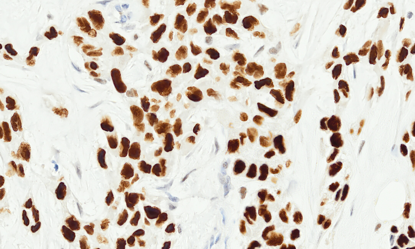
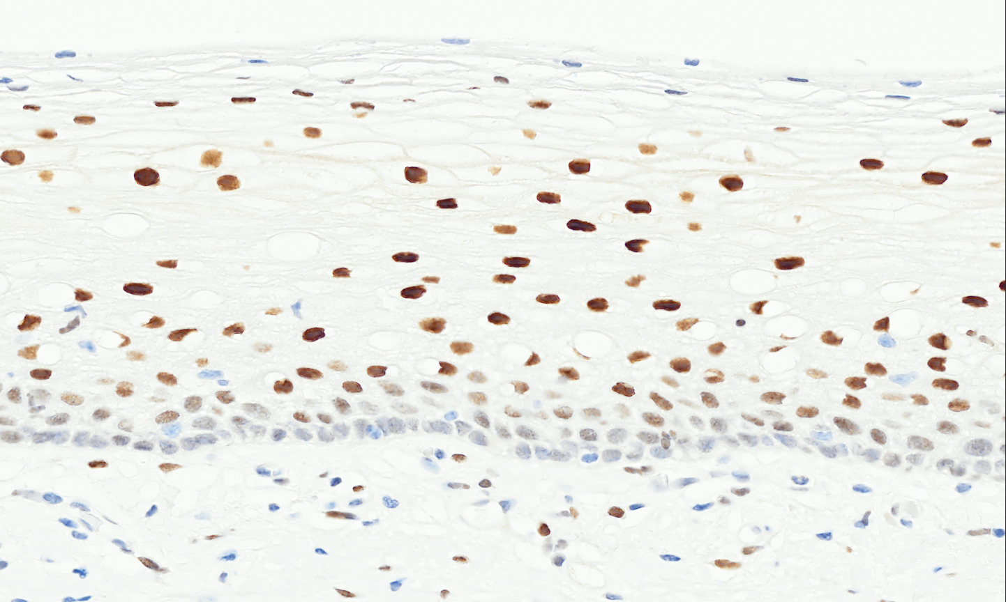
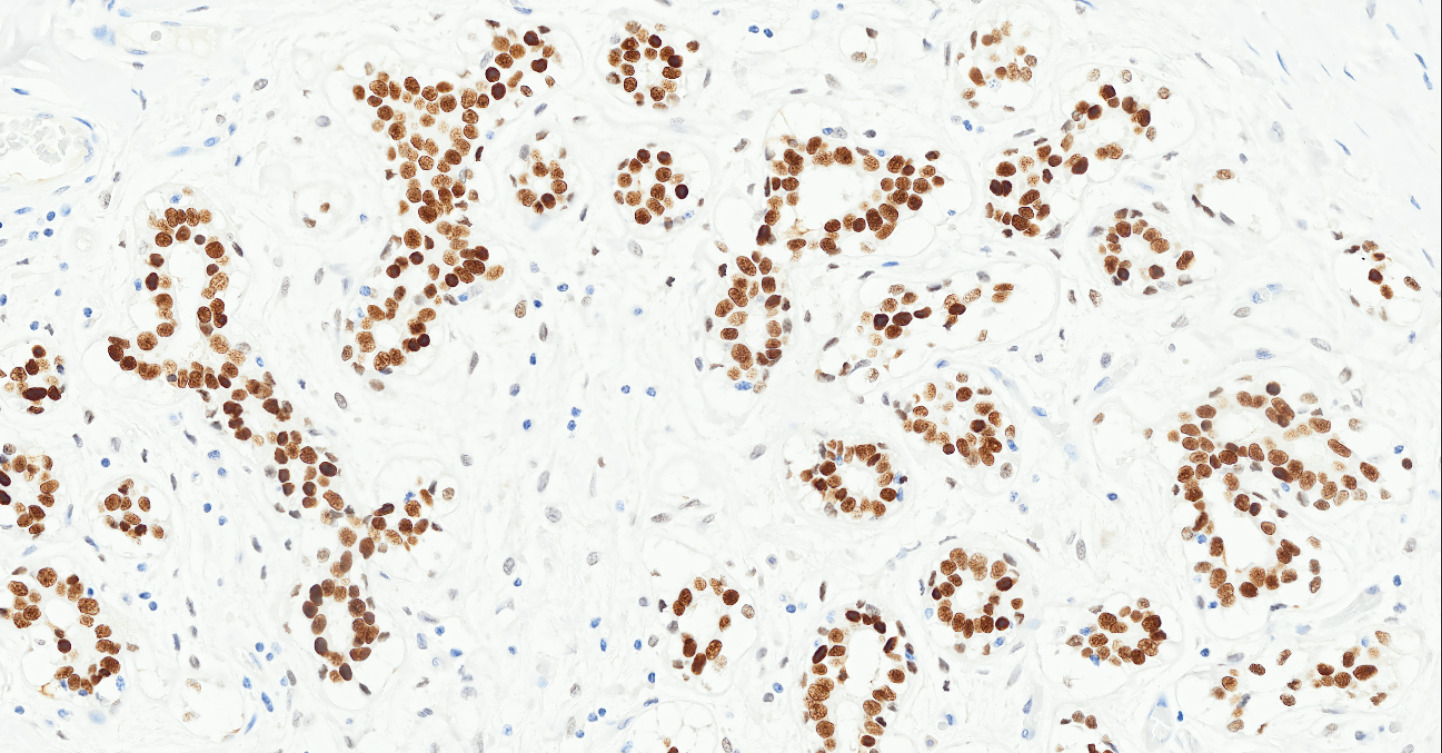
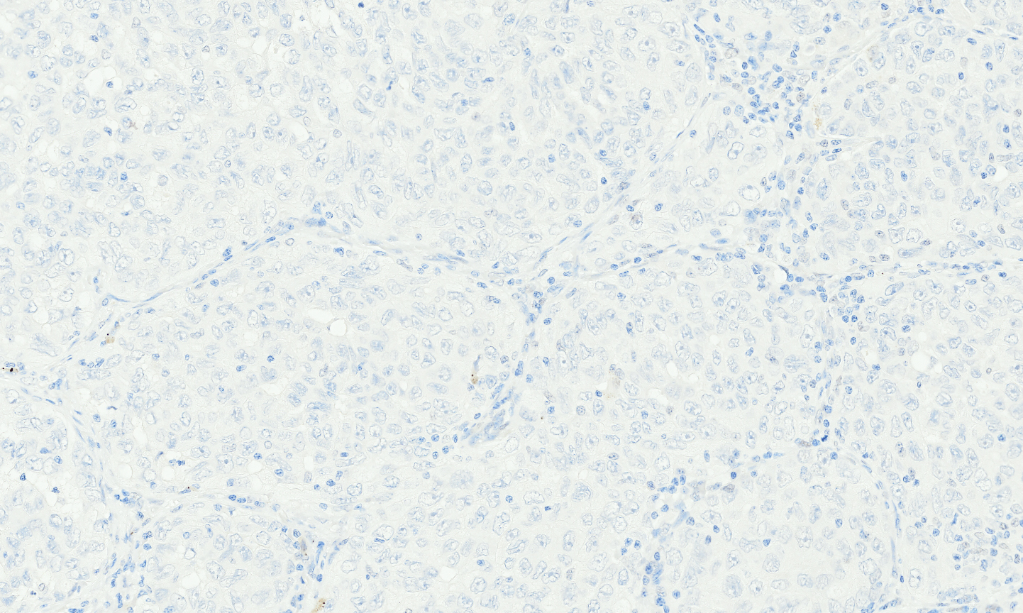
The Tissue-Tek Genie® anti- TRPS1 Rabbit Monoclonal Antibody [ZR382] is designed to qualitatively detect TRPS1 protein in formalin-fixed, paraffin-embedded (FFPE) human tissue specimen sections by immunohistochemistry (IHC) staining on the automated Tissue-Tek Genie® Advanced Staining System. This device functions as an aid for diagnosis and shall be used by a qualified pathologist with a panel of other antibodies to differentiate breast carcinoma, including triple negative subtypes, from cancer of unknown primary. Because of its higher sensitivity compared to established markers such as GATA3, GCDFP-15, Mammaglobin, and FOXC1, TRPS1 has recently gained recognition as a valuable adjunct IHC biomarker for diagnosing breast carcinoma. This is especially important in triple-negative breast cancers (TNBCs), where conventional markers often show low or absent expression. The frequent retention of TRPS1 expression in TNBCs makes it particularly useful for diagnosing primary tumors as well as identifying metastatic breast carcinoma.
Tissue-Tek Genie® anti-Tryptase
8372-M250: RTU, 1 cartridge, 250 tests; 1 unit
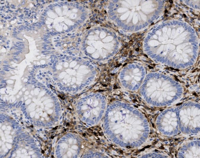
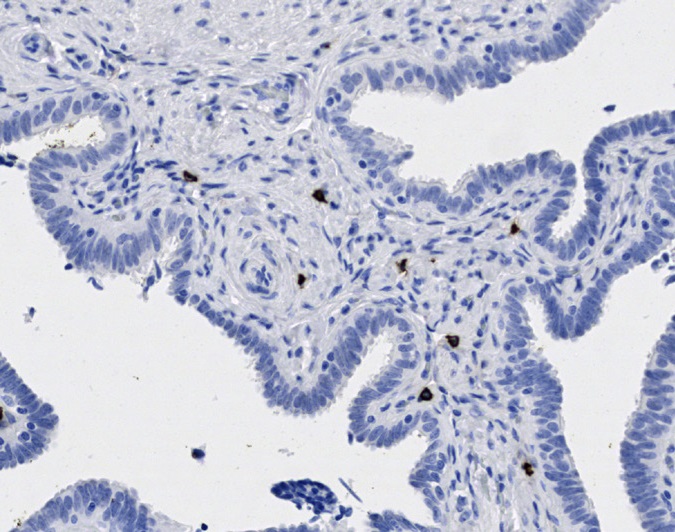
Tissue-Tek Genie® anti-Uroplakin II
8270-M250: RTU, 250 tests, 1 cartridge; 1 unit
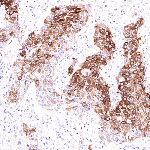
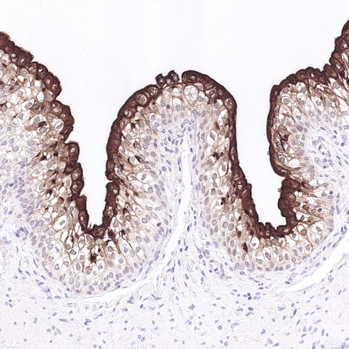
The Tissue-Tek Genie® anti-Uroplakin II Mouse Monoclonal Antibody [BC21] is designed to qualitatively detect Uroplakin II protein in formalin-fixed, paraffin-embedded (FFPE) human tissue specimen sections by immunohistochemistry (IHC) staining on the automated Tissue-Tek Genie® Advanced Staining System.
This device functions as an aid for diagnosis and shall be used by a qualified pathologist with a panel of other antibodies to differentiate urothelial carcinomas from other neoplasia.
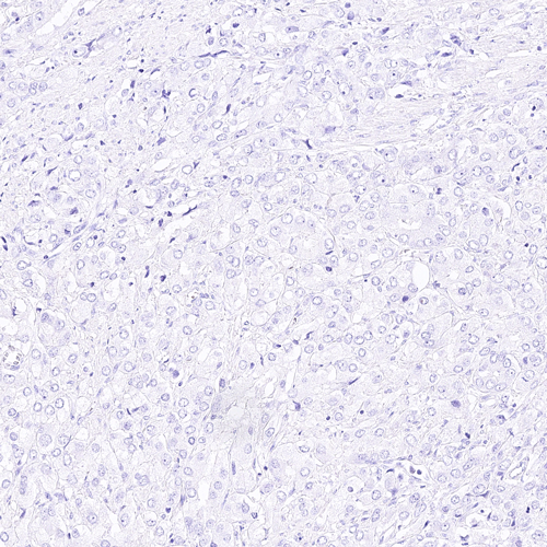
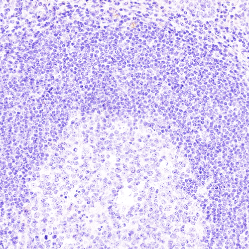
Tissue-Tek Genie® anti-Vimentin
8336-M250: RTU, 1 cartridge, 250 tests; 1 unit
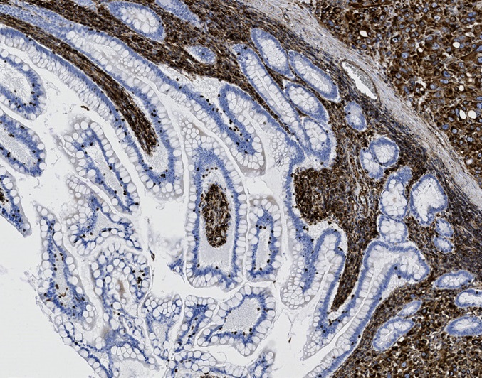
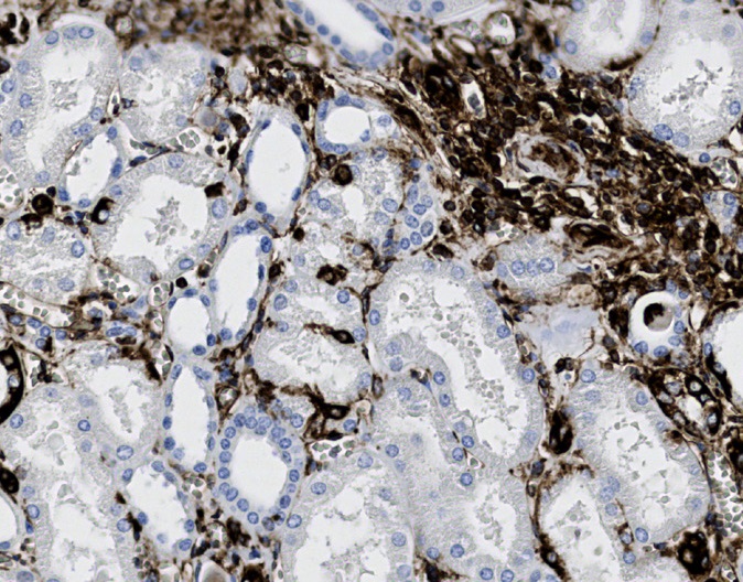
Tissue-Tek Genie® anti-WT1
8340-M250: RTU, 250 tests, 1 cartridge; 1 unit
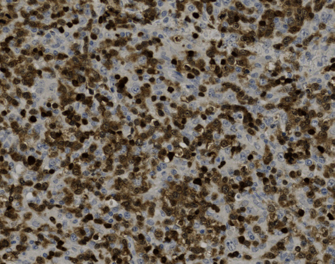
Tissue-Tek Genie® anti-ZAP70
8501-M250: RTU, 250 tests, 1 cartridge; 1 unit
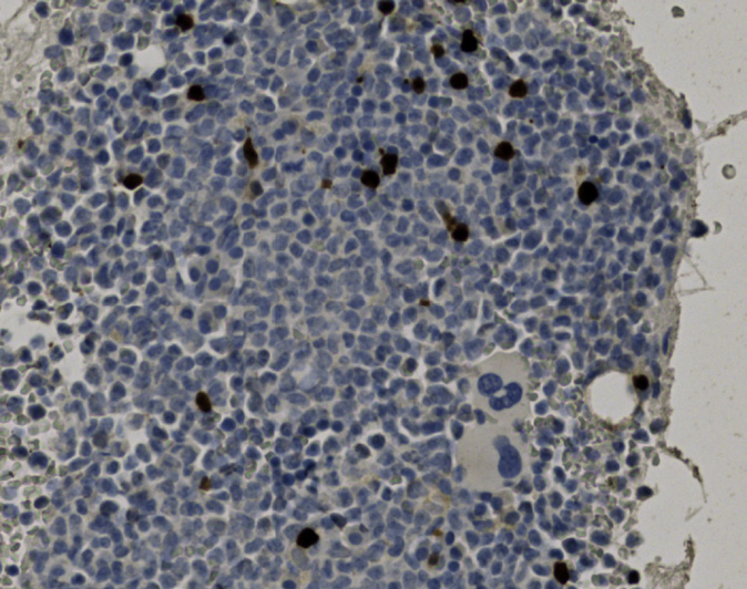
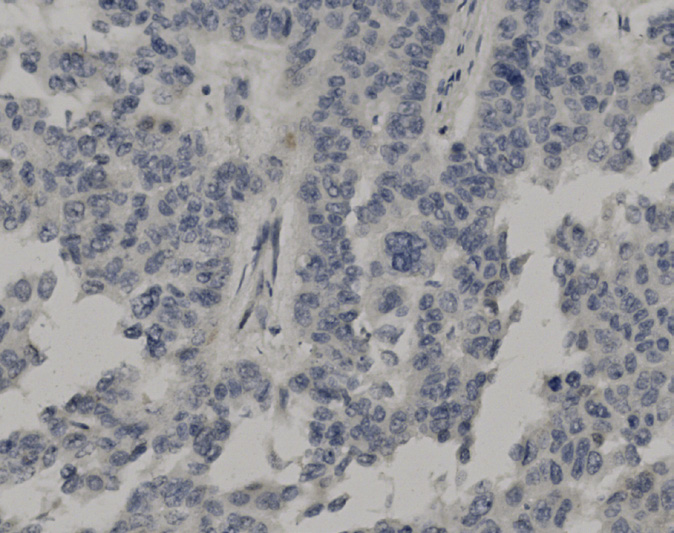
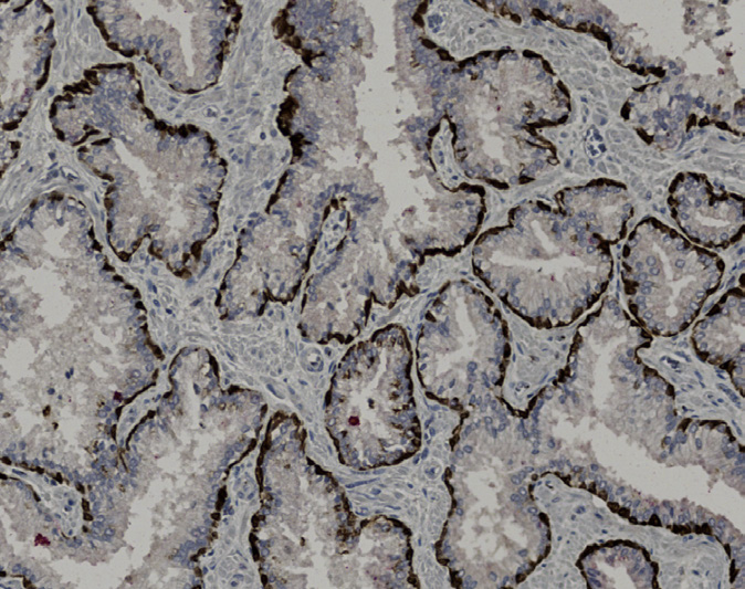
Tissue-Tek Genie® DUO anti-AMACR/p63/CK5/6 PIN Antibody Cocktail
8485-M100: RTU, 100 tests, 1 cartridge; 1 unit
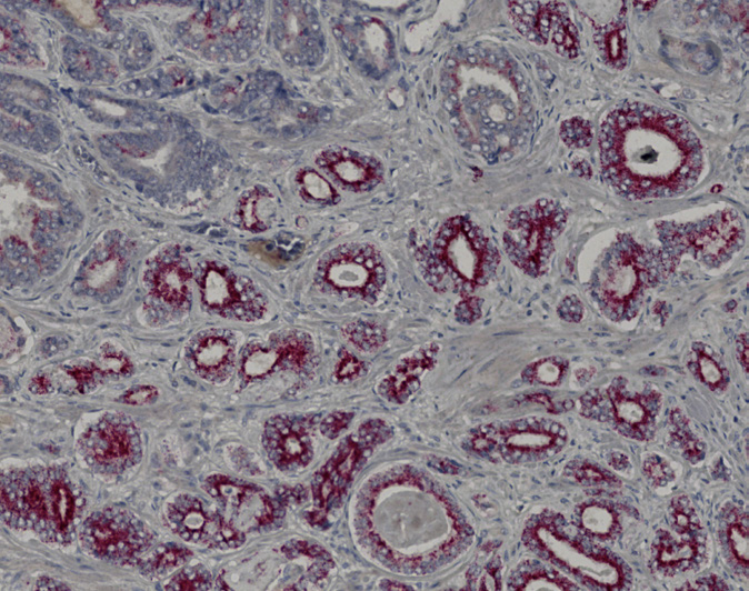
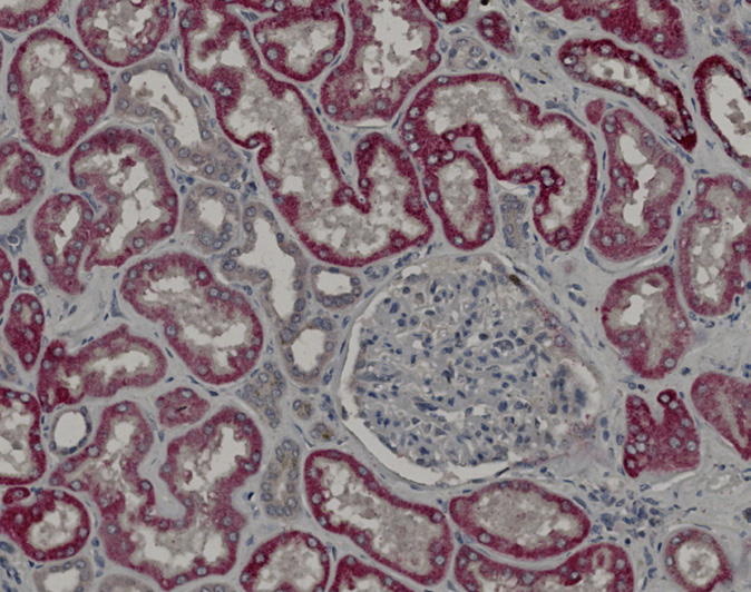
Tissue-Tek Genie® DUO anti-Melan-A / Ki67 Antibody Cocktail
8480-M100: RTU, 100 tests, 1 cartridge; 1 unit
Tissue-Tek Genie® DUO anti-p40 / Napsin A Antibody Cocktail
8487-M100: RTU, 100 tests, 1 cartridge; 1 unit
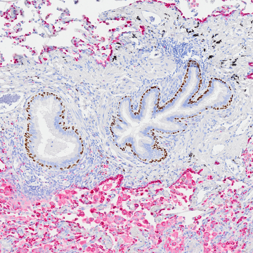
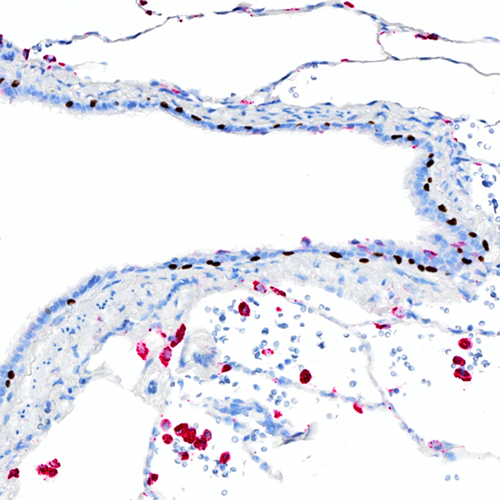
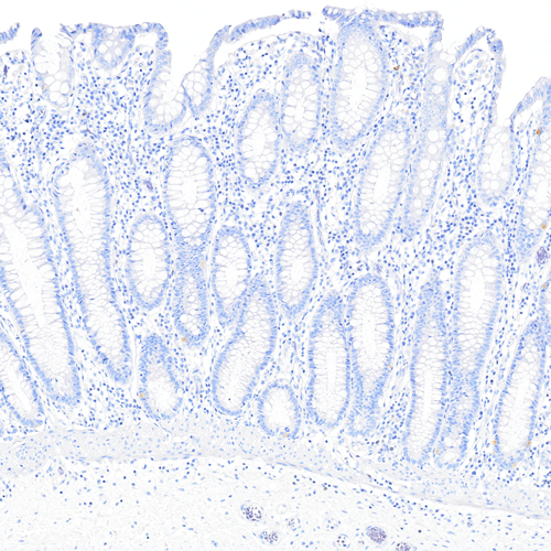
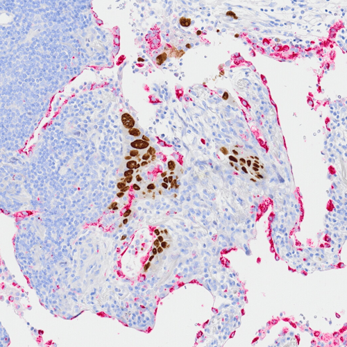
The Tissue-Tek Genie® DUO anti-p40 [BC28] / Napsin A [EP205] Antibody Cocktail is designed to qualitatively detect p40 and Napsin A proteins in formalin-fixed, paraffin-embedded (FFPE) human tissue specimen sections by immunohistochemistry (IHC) staining on the automated Tissue-Tek Genie® Advanced Staining System.
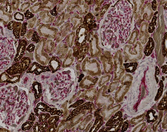
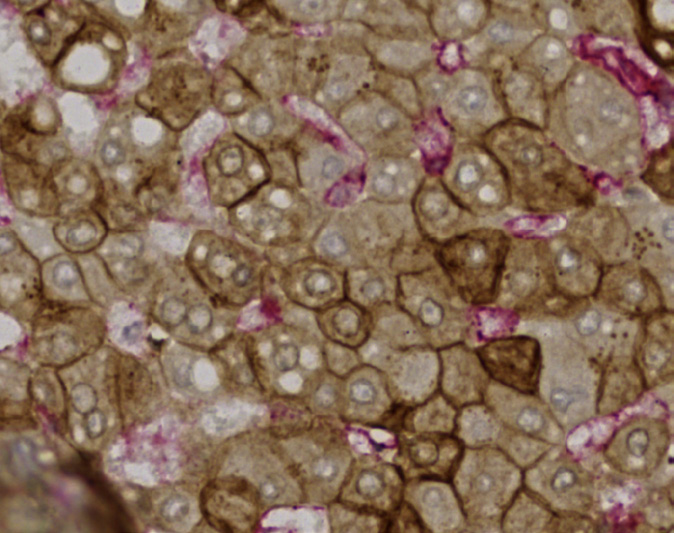
The Tissue-Tek Genie® DUO anti-p40 [BC28] / Napsin A [EP205] Antibody Cocktail utilizes the brown DAB chromogen for nuclear staining of p40 and the AP Red chromogen for cytoplasmic staining of Napsin A. When used with a panel of other antibodies, the antibody cocktail helps differentiate lung squamous cell carcinoma (most often p40 positive and Napsin A negative) from lung adenocarcinoma (most often p40 negative and Napsin A positive).
The DUO anti-p40 and Napsin A antibody cocktail labels p40 and Napsin A proteins in normal and neoplastic cells, and the component antibodies have a nuclear and cytoplasmic staining pattern, respectively.
Tissue-Tek Genie® DUO anti-Pan Cytokeratin/CD31 Antibody Cocktail
8484-M100: RTU, 100 tests, 1 cartridge; 1 unit
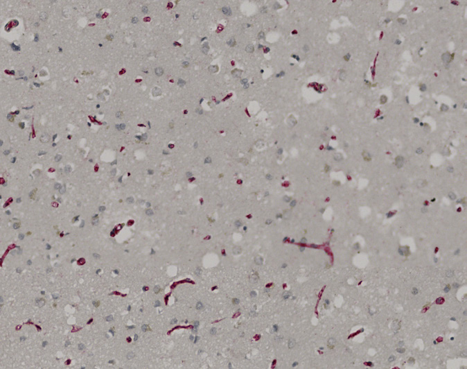
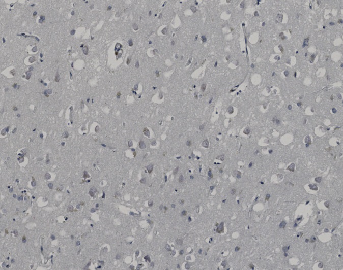
Tissue-Tek Genie® DUO Non-immune Mouse and Rabbit Ig Antibody Cocktail, Negative Control
8482-M250: RTU, 250 tests, 1 cartridge; 1 unit
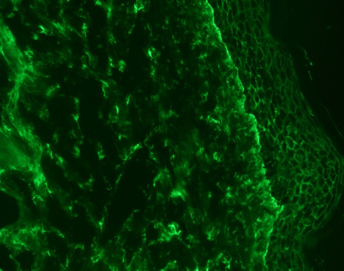
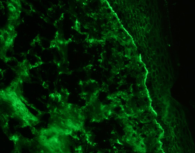
Tissue-Tek Genie® FITC anti-C3
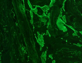
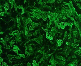
Tissue-Tek Genie® FITC anti-Fibrinogen
Tissue-Tek Genie® FITC anti-IgA
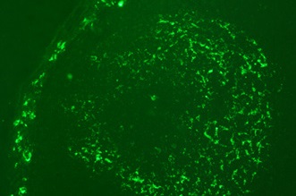
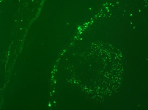
Tissue-Tek Genie® FITC anti-IgG
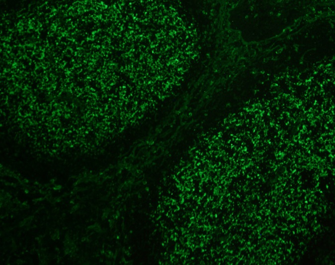
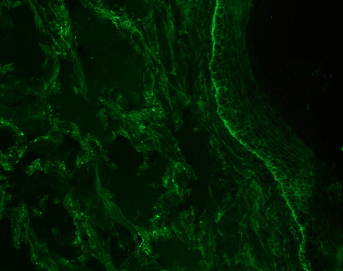
Tissue-Tek Genie® FITC anti-IgM
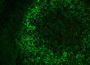
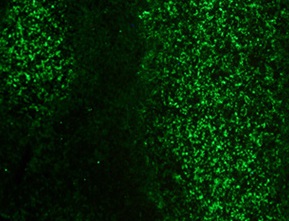
Tissue-Tek Genie® Pro Detection Kit, DAB

Product code and quantity
8826-K250: 6 cartridges, 250 tests, RTU; 1 kit
Description
The Tissue-Tek Genie Pro Detection Kit, DAB uses a non-biotin based system to detect and to visualize mouse or rabbit primary antibodies bound to antigens in Formalin-Fixed, Paraffin-Embedded (FFPE) specimen sections.
The detection system uses Protein and Peroxidase blocks to prevent unspecific binding of antibodies to antigens and reactions of endogenous peroxidase with downstream components of the detection system. After blocking, the detection system uses a Link solution that binds to the mouse or rabbit primary antibodies to form a complex with a horse radish peroxidase (HRP) conjugate which increases sensitivity.
The complex is then visualized with the addition of DAB. DAB Intensifier can be used to increase the intensity of
DAB staining.
Components
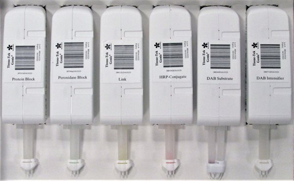
The detection system kit contains 6 cartridges, one cartridge each of the following:
• Tissue-Tek Genie® Protein Block, 250 tests
• Tissue-Tek Genie® Peroxidase Block, 250 tests
• Tissue-Tek Genie® Link, 250 tests
• Tissue-Tek Genie® Poly HRP-Conjugate, 250 tests
• Tissue-Tek Genie® DAB, 250 tests
• Tissue-Tek Genie® DAB Intensifier, 250 tests
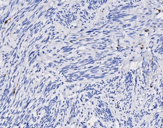
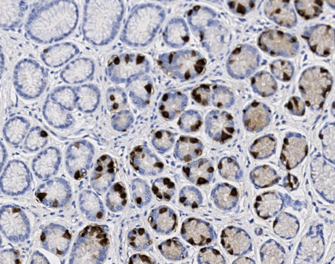
These 6 images display the exceptional sensitivity (high dynamic range) and superb specificity (no visible non-specific background staining) of the Tissue-Tek Genie Pro Detection Kit, DAB and selected Tissue-Tek Genie antibodies.
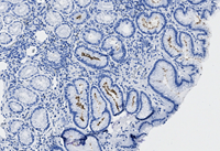
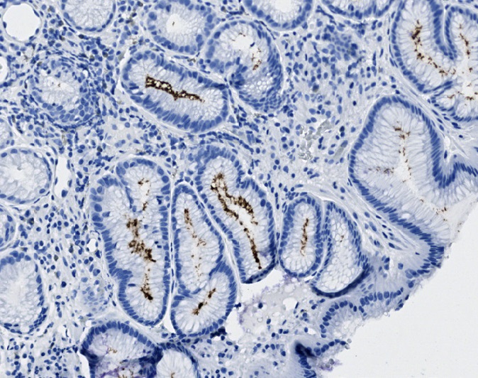
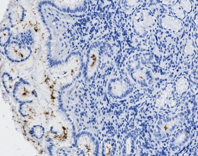
Positive Helicobacter pylori staining in crypts of gastric mucosa, no background staining.
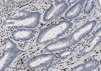
Negative staining of colon adenocarcinoma with loss of MLH1 expression, no background staining.
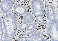
Negative staining of colon adenocarcinoma with loss of MSH2 expression, no background staining.
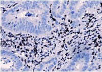
Negative staining of colon adenocarcinoma with loss of PMS2 expression, no background staining.
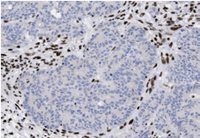
Negative staining of colon adenocarcinoma with loss of PMS2 expression, no background staining.
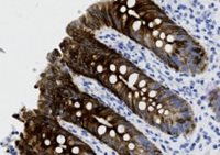
CK 19 staining in crypt cells of appendix with high dynamic range, no background staining.
The Tissue-Tek Genie Pro Detection Kit, DAB HRP staining steps provide standardization using RTU components and timing flexibility where needed.

Tissue-Tek Genie® Pro AP Red Detection KitTissue-Tek Genie® Pro AP Red Detection Kit
Product code and quantity
8836-K250: 4 cartridges, 250 tests, RTU; 1 kit
Description
The Tissue-Tek Genie Pro AP Red Detection Kit uses a non-biotin based system to detect and to visualize mouse and rabbit primary antibodies bound to antigens informalin-fixed, paraffin-embedded (FFPE) specimen sections.
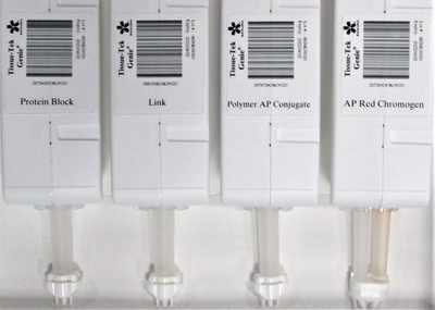
The detection system uses protein block to prevent nonspecific binding of antibodies to antigens with downstream components of the detection system. After blocking, the detection system uses a link solution that binds to the mouse and rabbit primary antibodies to form a complex with an alkaline phosphatase AP-Conjugate. The complex is then visualized with the addition of AP Red.
Components
The detection system kit contains 4 cartridges, one cartridge each of the following:
- Tissue-Tek Genie® Protein Block
- Tissue-Tek Genie® Link
- Tissue-Tek Genie® Poly AP-Conjugate
- Tissue-Tek Genie® AP Red
These 3 images display the exceptional sensitivity (high dynamic range) and superb specificity (no visible non-specific background staining) of the Tissue-Tek Genie Pro AP Red Detection Kit and selected Tissue-Tek Genie antibodies.
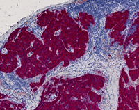
Strong pan melanoma staining of melanoma.
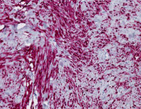
Melan-A staining of blue nevus.
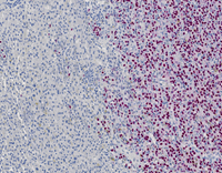
SOX10 staining of melanoma.
The Tissue-Tek Genie Pro AP Red Detection Kit, staining steps provide standardization using RTU components and timing flexibility where needed.

Tissue-Tek Genie® DUO Detection Kit
Product code and quantity
8837-K250: 6 cartridges, 250 tests, RTU; 1 kit
Tissue-Tek Genie® DUO Mouse-DAB/Rabbit-AP Red Dual Detection Kit
The Tissue-Tek Genie® DUO Mouse-DAB/Rabbit-AP Red Dual Detection Kit uses a non-biotin-based system to detect and to visualize mouse and rabbit primary antibodies bound to antigens in FFPE specimen sections.
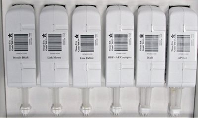
The detection system uses a protein block to prevent nonspecific binding of antibodies to antigens with downstream components of the detection system. After blocking, mouse and rabbit primary antibodies are applied on the tissue sections simultaneously using prefilled capsules or cartridges. Subsequently, the detection system sequentially applies the Link Mouse solution that binds to mouse primary antibodies and then the Link Rabbit solution that binds to rabbit primary antibodies. This is followed by an application of a cocktail containing a horseradish peroxidase (HRP) conjugate for mouse and an alkaline phosphatase AP-Conjugate for rabbit. The resulting complex is visualized by sequentially applying the diaminobenzidine (DAB) substrate-chromogen solution, and then by applying the AP Red substrate-chromogen solution, which create colored precipitates at the location of the antigens.

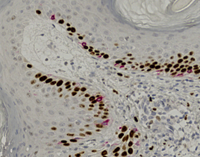
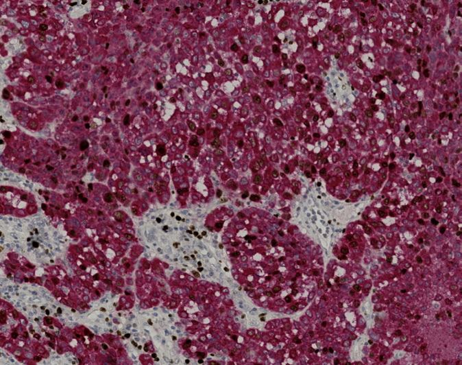
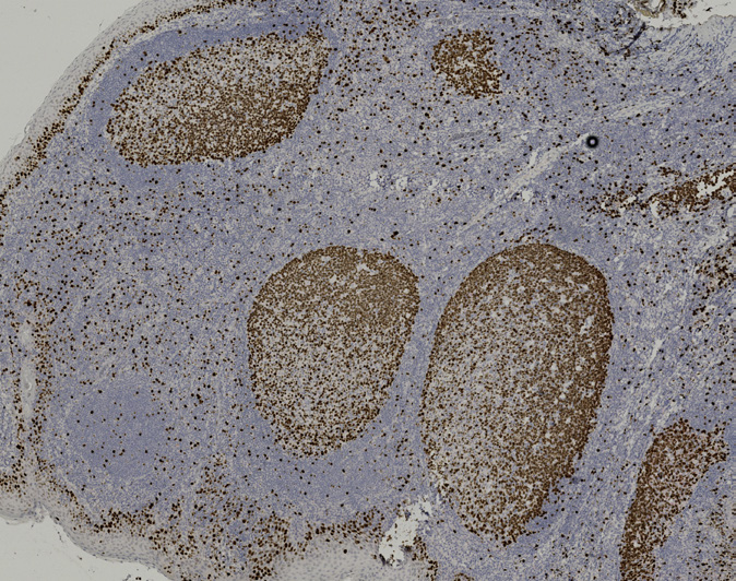
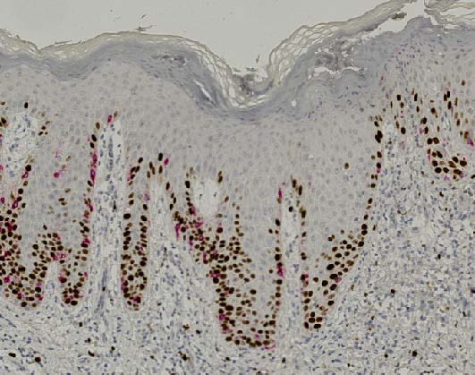
Tissue-Tek Genie® DUO anti-Melan-A [EP43] /Ki67 [GM010] Antibody Cocktail.
Note that the benign melanocytes are not proliferatively active.
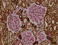
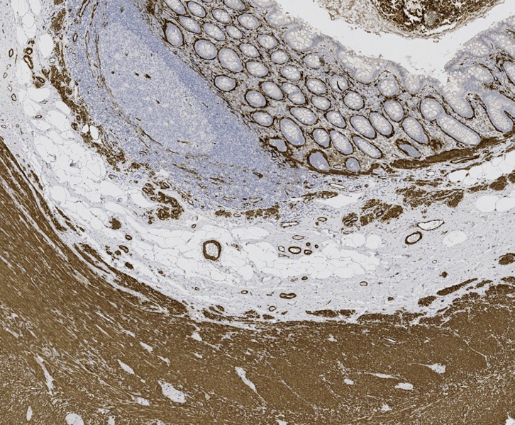
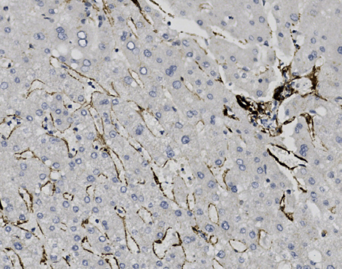
Tissue-Tek Genie® DUO anti-Pan Cytokeratin [AE1/AE3/DC10] / CD31[RM247] Antibody Cocktail.
The combination of these two markers on a single slide is particularly helpful for identifying small foci of carcinoma within lymphatics.
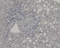
Tissue-Tek Genie® DUO Non-immune Mouse and Rabbit Ig Antibody Cocktail.
Tissue-Tek Genie® Pro Antibody Diluent
Product code and quantity
8866-G004: RTU, 4 bottles, 100 mL each; 1 case
Description
Optimal staining performance during immunohistochemistry (IHC) is achieved through dilution of the primary antibodies. This dilution process involves mixing concentrated antibody with Tissue-Tek Genie® Pro Antibody Diluent to an optimal dilution determined by the user. The diluted antibody can then be filled into a Tissue-Tek Genie® User-Fillable Capsule, Tissue-Tek Genie® User Fillable Cartridge, or be applied by hand during manual application mode on the Tissue-Tek Genie® Advanced Staining System.
Tissue-Tek Genie® Hematoxylin
Product code and quantity
8830-M250: RTU, 250 tests, 1 cartridge: 1 unit
Description
The Tissue-Tek Genie® Hematoxylin is designed to counterstain cellular nuclei in formalinfixed, paraffin embedded (FFPE) human specimen sections after immunohistochemistry (IHC) staining of specific protein targets or chromogenic in situ hybridization (CISH) of specific DNA or RNA targets using the automated Tissue-Tek Genie® Advanced Staining System.
Tissue-Tek Genie® Citrate Antigen Retrieval Solution
Product code and quantity
8742-G001: RTU, 1 bottle, 3.8 Liters; 1 unit
Description
Tissue-Tek Genie® Citrate Antigen Retrieval Solution is designed for heat-induced epitope retrieval (HIER) of specimen sections during immunohistochemical (IHC) staining on the Tissue-Tek Genie® Advanced Staining System.
Tissue fixation involves creating crosslinks between protein residues in the tissue. This fixation process preserves the tissue, but also denatures many antigens and makes antigen sites within the tissue less accessible to antibodies. The Tissue-Tek Genie Citrate Antigen Retrieval Solution is applied to the specimen section and heated on the Tissue-Tek Genie Advanced Staining System. This process breaks the crosslinks formed during fixation and allows proteins to renature which allows greater accessibility of antigenic sites to antibodies.
Tissue-Tek Genie® Dewax Solution
Product code and quantity
8865-G001: RTU, 1 bottle, 3.8 Liters; 1 unit
Description
Tissue-Tek Genie® Dewax Solution is designed to remove paraffin wax from paraffin embedded tissue sections on the Tissue-Tek Genie® Advanced Staining System.
Formalin-fixed, paraffin embedded (FFPE) specimen sections mounted on microscope slides are treated with Tissue-Tek Genie Dewax Solution on the Tissue-Tek Genie Advanced Staining System prior to the immunohistochemistry and/or in situ hybridization staining processes. Specimen sections in the presence of Tissue-Tek Genie Dewax Solution are gently heated for optimized removal of paraffin.
Tissue-Tek Genie® High pH Antigen Retrieval Solution
Product code and quantity
8744-G001: RTU, 1 bottle, 3.8 Liters; 1 unit
Description
Tissue-Tek Genie® High pH Antigen Retrieval Solution is designed for heat-induced epitope retrieval (HIER) of specimen sections during immunohistochemical (IHC) staining on the Tissue-Tek Genie® Advanced Staining System.
Tissue-Tek Genie® Wash Buffer Solution
Product code and quantity
8874-G004: RTU, 4 bottles, 3.8 Liters each: 1 case
Description
Tissue-Tek Genie® Wash Buffer Solution is designed to remove residual reagents from tissue sections during immunohistochemical (IHC) staining on the Tissue-Tek Genie® Advanced Staining System.
The Tissue-Tek Genie Wash Buffer Solution is a bulk reagent that is used between incubation steps during IHC or ISH staining processes. The Tissue-Tek Genie Wash Buffer Solution serves as a rinsing agent for the removal of residual reagents that remain on the tissue sections from the previous protocol step.
Tissue-Tek Genie® Proteinase K
Product code and quantity
9811-M100: RTU, 100 tests, 1 cartridge; 1 unit
Description
The Tissue-Tek Genie® Proteinase K is intended for the proteolytic digestion of formalin-fixed, paraffin embedded (FFPE) human tissue sections during immuno-histochemistry (IHC) staining of specific protein targets or chromogenic in situ hybridization (CISH) staining of specific DNA or RNA targets on the automated Tissue-Tek Genie® Advanced Staining System.
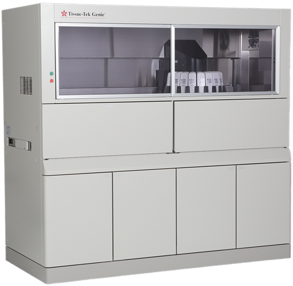
Need more information?
Our team members are ready to support your laboratory needs or answer any questions you may have.
Contact