Clone: EP43/GM010
Host clonality: mouse and rabbit monoclonal
Control tissue: melanoma, skin
Staining pattern: cytoplasmic for Melan-A and nuclear for Ki67
Regulatory status: IVD, FDA Class I
|
Product code:
|
8480-C010: RTU, 10 capsules; 1 pack
8480-M100: RTU, 100 tests, 1 cartridge; 1 unit
|
Melan-A, also known as Melanoma Antigen Recognized by T-cell 1 (MART-1), is a protein associated with endoplasmic reticulum and melanosomes and is expressed in melanocytes. Melan-A is expressed in all normal melanocytes and is detected in 80-100% of melanomas. Ki67 is a proliferation-associated nuclear protein is expressed during all active phases of the cell cycle and is absent in resting cells.

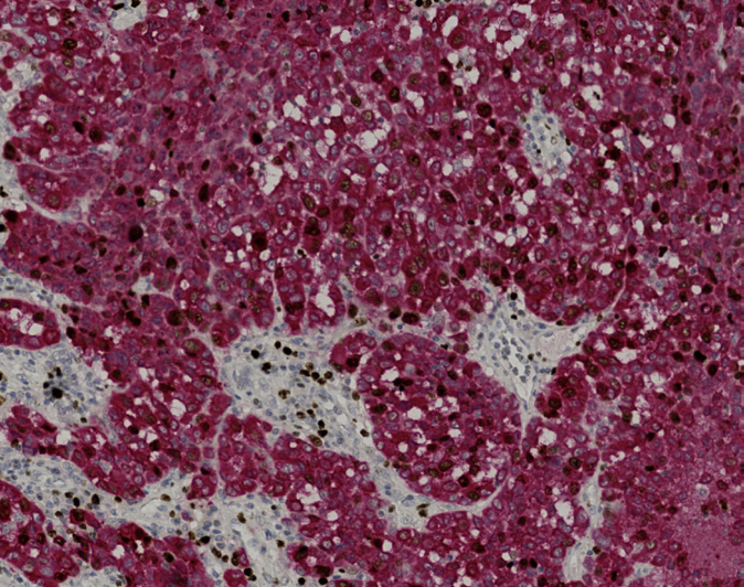
The neoplastic cells of this melanoma show strong cytoplasmic staining for Melan-A in red, with a significant fraction showing nuclear Ki67 reactivity in brown.
.jpg)
No staining reaction for Melan-A is observed in this reactive tonsil, while an extensive, strong nuclear staining reaction is present among the proliferating germinal center lymphocytes and the basal cells of the squamous epithelium.
.jpg)
The melanocytes in the basal epidermis show moderate to strong cytoplasmic reactivity for Melan-A in red. The proliferating basal keratinocytes show moderate to strong Ki67 nuclear reactivity in brown.