Clone: EP239
Host clonality: rabbit monoclonal
Control tissue: tonsil, lymph-node, selective T-cell lymphomas
Staining pattern: cytoplasmic
Regulatory status: IVD, FDA Class I
|
Product code:
|
8287-C010: RTU, 10 capsules; 1 pack
8287-M250: RTU, 250 tests, 1 cartridge; 1 unit
|
Programmed-death ligand 1 (PD-1) is expressed by germinal center-associated helper T-cells and inhibits T-cell activity. PD-1 is expressed on T-cells, B-cells, and monocytes during activation. PD-1 positivity has been found in angioimmunoblastic lymphoma, primary cutaneous pleomorphic T-cell lymphoma, and nodular lymphocyte- predominant Hodgkin lymphoma but not other subtypes of T-cell and B-cell non-Hodgkin lymphomas or classic Hodgkin lymphomas.

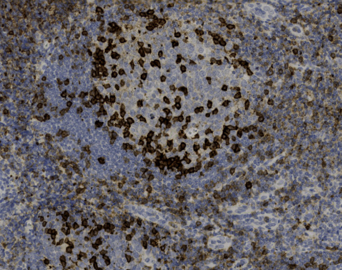
Variable reactivity is seen for PD-1 in this tonsil specimen with the darkest cytoplasmic reactivity in cells within the follicle.
.jpg)
Lymphocytes of the colonic lamina propria demonstrate variable, weak to strong, cytoplasmic expression of PD-1.
.jpg)
Pancreatic acini and ducts, as well as islet cells are all negative PD-1.